Radial glial dependent and independent dynamics of interneuronal migration in the developing cerebral cortex
- PMID: 17726524
- PMCID: PMC1950908
- DOI: 10.1371/journal.pone.0000794
Radial glial dependent and independent dynamics of interneuronal migration in the developing cerebral cortex
Abstract
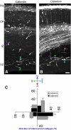
Interneurons originating from the ganglionic eminence migrate tangentially into the developing cerebral wall as they navigate to their distinct positions in the cerebral cortex. Compromised connectivity and differentiation of interneurons are thought to be an underlying cause in the emergence of neurodevelopmental disorders such as schizophrenia. Previously, it was suggested that tangential migration of interneurons occurs in a radial glia independent manner. Here, using simultaneous imaging of genetically defined populations of interneurons and radial glia, we demonstrate that dynamic interactions with radial glia can potentially influence the trajectory of interneuronal migration and thus the positioning of interneurons in cerebral cortex. Furthermore, there is extensive local interneuronal migration in tangential direction opposite to that of pallial orientation (i.e., in a medial to lateral direction from cortex to ganglionic eminence) all across the cerebral wall. This counter migration of interneurons may be essential to locally position interneurons once they invade the developing cerebral wall from the ganglionic eminence. Together, these observations suggest that interactions with radial glial scaffold and localized migration within the expanding cerebral wall may play essential roles in the guidance and placement of interneurons in the developing cerebral cortex.
Conflict of interest statement
Figures


Similar articles
-
Modes of neuronal migration in the developing cerebral cortex.Nat Rev Neurosci. 2002 Jun;3(6):423-32. doi: 10.1038/nrn845. Nat Rev Neurosci. 2002. PMID: 12042877 Review.
-
Neuronal migration in the developing cerebral cortex: observations based on real-time imaging.Cereb Cortex. 2003 Jun;13(6):607-11. doi: 10.1093/cercor/13.6.607. Cereb Cortex. 2003. PMID: 12764035 Review.
-
Multidirectional and multizonal tangential migration of GABAergic interneurons in the developing cerebral cortex.Development. 2006 Jun;133(11):2167-76. doi: 10.1242/dev.02382. Epub 2006 May 3. Development. 2006. PMID: 16672340
-
Transient maternal hypothyroxinemia at onset of corticogenesis alters tangential migration of medial ganglionic eminence-derived neurons.Eur J Neurosci. 2005 Aug;22(3):541-51. doi: 10.1111/j.1460-9568.2005.04243.x. Eur J Neurosci. 2005. PMID: 16101736
-
Connexin 43 mediates the tangential to radial migratory switch in ventrally derived cortical interneurons.J Neurosci. 2010 May 19;30(20):7072-7. doi: 10.1523/JNEUROSCI.5728-09.2010. J Neurosci. 2010. PMID: 20484649 Free PMC article.
Cited by
-
Dynamics of the leading process, nucleus, and Golgi apparatus of migrating cortical interneurons in living mouse embryos.Proc Natl Acad Sci U S A. 2012 Oct 9;109(41):16737-42. doi: 10.1073/pnas.1209166109. Epub 2012 Sep 24. Proc Natl Acad Sci U S A. 2012. PMID: 23010922 Free PMC article.
-
The essential role of primary cilia in cerebral cortical development and disorders.Curr Top Dev Biol. 2021;142:99-146. doi: 10.1016/bs.ctdb.2020.11.003. Epub 2021 Jan 25. Curr Top Dev Biol. 2021. PMID: 33706927 Free PMC article. Review.
-
Coordinating cerebral cortical construction and connectivity: Unifying influence of radial progenitors.Neuron. 2022 Apr 6;110(7):1100-1115. doi: 10.1016/j.neuron.2022.01.034. Epub 2022 Feb 24. Neuron. 2022. PMID: 35216663 Free PMC article. Review.
-
Random walk behavior of migrating cortical interneurons in the marginal zone: time-lapse analysis in flat-mount cortex.J Neurosci. 2009 Feb 4;29(5):1300-11. doi: 10.1523/JNEUROSCI.5446-08.2009. J Neurosci. 2009. PMID: 19193877 Free PMC article.
-
Transcriptional dysregulation of neocortical circuit assembly in ASD.Int Rev Neurobiol. 2013;113:167-205. doi: 10.1016/B978-0-12-418700-9.00006-X. Int Rev Neurobiol. 2013. PMID: 24290386 Free PMC article. Review.
References
-
- Marin O, Rubenstein JL. Cell migration in the forebrain. Annu Rev Neurosci. 2003;26:441–83. - PubMed
-
- Miyata T, Kawaguchi A, Okano H, Ogawa M. Asymmetric inheritance of radial glial fibers by cortical neurons. Neuron. 2001;31:727–41. - PubMed
-
- Nadarajah B, Brunstrom JE, Grutzendler J, Wong RO, Pearlman AL. Two modes of radial migration in early development of the cerebral cortex. Nat Neurosci. 2001;4:143–150. - PubMed
-
- Parnavelas JG, Nadarajah B. Radial glial cells. are they really glia? Neuron. 2001;31:881–4. - PubMed
-
- Anderson SA, Eisenstat DD, Shi L, Rubenstein JL. Interneuron migration from basal forebrain to neocortex: dependence on Dlx genes. Science. 1997;278:474–476. - PubMed
Publication types
MeSH terms
Grants and funding
LinkOut - more resources
Full Text Sources
Molecular Biology Databases

