. 2007 Aug 29;2(8):e799.
doi: 10.1371/journal.pone.0000799.

Phage-Antibiotic Synergy (PAS): beta-lactam and quinolone antibiotics stimulate virulent phage growth

Affiliations

Phage-Antibiotic Synergy (PAS): beta-lactam and quinolone antibiotics stimulate virulent phage growth

André M Comeau et al. PLoS One. .

Abstract

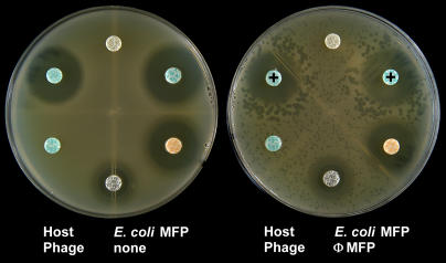
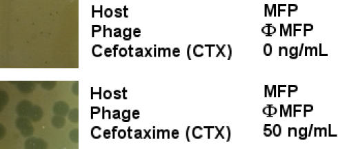
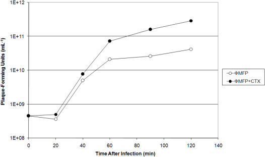
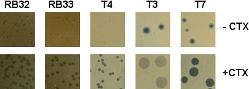
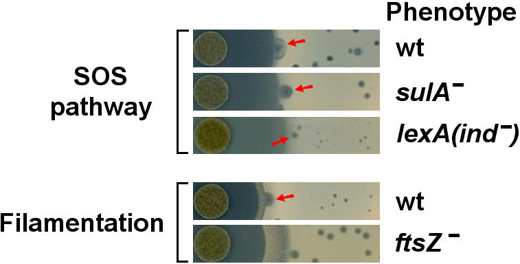
Although the multiplication of bacteriophages (phages) has a substantial impact on the biosphere, comparatively little is known about how the external environment affects phage production. Here we report that sub-lethal concentrations of certain antibiotics can substantially stimulate the host bacterial cell's production of some virulent phage. For example, a low dosage of cefotaxime, a cephalosporin, increased an uropathogenic Escherichia coli strain's production of the phage PhiMFP by more than 7-fold. We name this phenomenon Phage-Antibiotic Synergy (PAS). A related effect was observed in diverse host-phage systems, including the T4-like phages, with beta-lactam and quinolone antibiotics, as well as mitomycin C. A common characteristic of these antibiotics is that they inhibit bacterial cell division and trigger the SOS system. We therefore examined the PAS effect within the context of the bacterial SOS and filamentation responses. We found that the PAS effect appears SOS-independent and is primarily a consequence of cellular filamentation; it is mimicked by cells that constitutively filament. The fact that completely unrelated phages manifest this phenomenon suggests that it confers an important and general advantage to the phages.


Conflict of interest statement

Competing Interests: Three of the paper's authors (F.T., M.-F.P., and H.M.K.) are authors of a patent application (PCT/IB2007/000880), filed under the Patent Cooperation Treaty (PCT), involving some of the work presented in this paper.

Figures

Figure 1
Figure 1. The PAS effect of phage ΦMFP on E. coli MFP on Luria-Bertani agar plates.
Only disks containing the β-lactam antibiotics aztreonam and cefixime (indicated by “+” symbols) produced large phage plaques in their proximity. Gentamicin and tetracycline gave no PAS effect. This host strain was resistant to both amoxicillin and trimethoprim/sulfamethoxazole. Note the absence of phages on the left-hand control plate indicating the lack of prophage induction.
Figure 2
Figure 2. Plaque sizes of phage ΦMFP on E. coli MFP with and without 50 ng/mL of cefotaxime (CTX) in Luria agar plates.
Figure 3
Figure 3. Increase in phage titer in the presence of the cephalosporin cefotaxime (CTX).
E. coli strain MFP was infected with phage ΦMFP in Luria liquid medium supplemented at the time of infection with 20 ng/mL of cefotaxime, or left untreated. The multiplicity of infection was ∼5. Chloroform was added at various times after infection to lyse the infected cells.
Figure 4
Figure 4. Plaque sizes of various coliphages with and without cefotaxime (CTX) in Luria agar plates.
Phages RB32 and RB33 were grown on E. coli strain MFP (50 ng/mL CTX) at 37°C; and T4, T3 and T7 were grown on E. coli strain AS19 (30 ng/mL CTX) at 25°C. All plaques were photographed at identical magnifications.
Figure 5
Figure 5. The PAS effect of phage T4 on various E. coli SOS and filamentation mutant strains.
T4 was grown on E. coli sulA-inactivated and lexA non-inducible mutant strains (defective SOS systems), as well as an ftsZ-inactivated mutant strain (non-antibiotic induced filamentation), in the presence of disks of cefotaxime (CTX). Isogenic wild-type strains (wt) are also included and representative plaques demonstrating the PAS effect are indicated by red arrows. All plates were photographed at identical magnifications.

Similar articles

Cited by

References

    1. Karam J, editor. Washington DC: American Society for Microbiology Press; 1994. Molecular biology of bacteriophage T4. p. 633.
    1. Oppenheim AB, Kobiler O, Stavans J, Court DL, Adhya S. Switches in bacteriophage lambda development. Annu Rev Genet. 2005;39:409–429. - PubMed
    1. Gottfredsson M, Erlendsdóttir H, Sigfússon A, Gudmundsson S. Characteristics and dynamics of bacterial populations during postantibiotic effect determined by flow cytometry. Antimicrob Agents Chemother. 1998;42:1005–1011. - PMC - PubMed
    1. Piddock LJV, Walters RN, Diver JM. Correlation of quinolone MIC and inhibition of DNA, RNA, and protein synthesis and induction of the SOS response in Escherichia coli. Antimicrob Agents Chemother. 1990;34:2331–2336. - PMC - PubMed
    1. Ackermann H-W. Bacteriophage observations and evolution. Res Microbiol. 2003;154:245–251. - PubMed

Publication types

MeSH terms