

Kinetic analysis of Yersinia pestis DNA adenine methyltransferase activity using a hemimethylated molecular break light oligonucleotide
- PMID: 17726531
- PMCID: PMC1949145
- DOI: 10.1371/journal.pone.0000801
Kinetic analysis of Yersinia pestis DNA adenine methyltransferase activity using a hemimethylated molecular break light oligonucleotide
Abstract
Background: DNA adenine methylation plays an important role in several critical bacterial processes including mismatch repair, the timing of DNA replication and the transcriptional control of gene expression. The dependence of bacterial virulence on DNA adenine methyltransferase (Dam) has led to the proposal that selective Dam inhibitors might function as broad spectrum antibiotics.
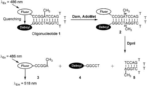
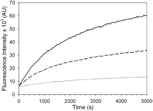
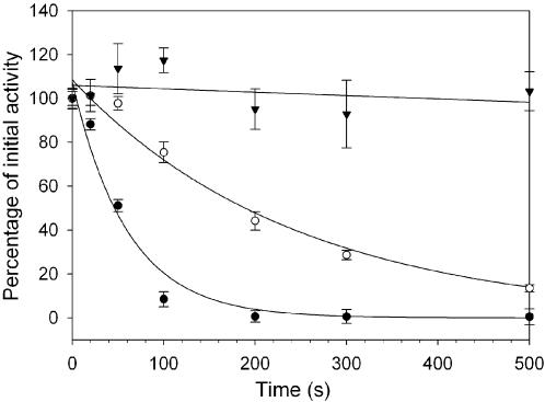
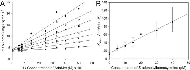
Methodology/principal findings: Herein we report the expression and purification of Yersinia pestis Dam and the development of a continuous fluorescence based assay for DNA adenine methyltransferase activity that is suitable for determining the kinetic parameters of the enzyme and for high throughput screening against potential Dam inhibitors. The assay utilised a hemimethylated break light oligonucleotide substrate containing a GATC methylation site. When this substrate was fully methylated by Dam, it became a substrate for the restriction enzyme DpnI, resulting in separation of fluorophore (fluorescein) and quencher (dabcyl) and therefore an increase in fluorescence. The assays were monitored in real time using a fluorescence microplate reader in 96 well format and were used for the kinetic characterisation of Yersinia pestis Dam, its substrates and the known Dam inhibitor, S-adenosylhomocysteine. The assay has been validated for high throughput screening, giving a Z-factor of 0.71+/-0.07 indicating that it is a sensitive assay for the identification of inhibitors.
Conclusions/significance: The assay is therefore suitable for high throughput screening for inhibitors of DNA adenine methyltransferases and the kinetic characterisation of the inhibition.
Conflict of interest statement
Figures
Similar articles
-
Inhibition of Yersinia pestis DNA adenine methyltransferase in vitro by a stibonic acid compound: identification of a potential novel class of antimicrobial agents.Br J Pharmacol. 2013 Jan;168(1):172-88. doi: 10.1111/j.1476-5381.2012.02134.x. Br J Pharmacol. 2013. PMID: 22889062 Free PMC article.
-
[Comparison of specific recognition sites of adenine and cytosine DNA-methylase of Yersinia Pestis EV 76 C dam and dcm by Escherichia coli methylases].Biokhimiia. 1984 Oct;49(10):1594-7. Biokhimiia. 1984. PMID: 6097301 Russian.
-
Direct and continuous fluorescence-based measurements of Pyrococcus horikoshii DNA N-6 adenine methyltransferase activity.Anal Biochem. 2011 Nov 15;418(2):204-12. doi: 10.1016/j.ab.2011.07.023. Epub 2011 Jul 27. Anal Biochem. 2011. PMID: 21839719
-
Roles of DNA adenine methylation in host-pathogen interactions: mismatch repair, transcriptional regulation, and more.FEMS Microbiol Rev. 2009 May;33(3):488-503. doi: 10.1111/j.1574-6976.2008.00159.x. Epub 2009 Jan 19. FEMS Microbiol Rev. 2009. PMID: 19175412 Free PMC article. Review.
-
[The DNA-methylation state regulates virulence and stress response of Salmonella].C R Biol. 2008 Sep;331(9):648-54. doi: 10.1016/j.crvi.2008.06.002. Epub 2008 Jul 2. C R Biol. 2008. PMID: 18722983 Review. French.
Cited by
-
A real-time assay for CpG-specific cytosine-C5 methyltransferase activity.Nucleic Acids Res. 2010 May;38(9):e107. doi: 10.1093/nar/gkq047. Epub 2010 Feb 5. Nucleic Acids Res. 2010. PMID: 20139415 Free PMC article.
-
Inhibition of Yersinia pestis DNA adenine methyltransferase in vitro by a stibonic acid compound: identification of a potential novel class of antimicrobial agents.Br J Pharmacol. 2013 Jan;168(1):172-88. doi: 10.1111/j.1476-5381.2012.02134.x. Br J Pharmacol. 2013. PMID: 22889062 Free PMC article.
References
-
- Jeltsch A. Beyond Watson and Crick: DNA methylation and molecular enzymology of DNA methyltransferases. Chembiochem. 2002;3:275–293. - PubMed
-
- Dryden DTF. 1999. S-Adenosylmethionine Dependent Methyltransferases: Structures and Functions: World Scientific, London. pp. 283–340.
-
- Geier GE, Modrich P. Recognition Sequence of the Dam Methylase of E-Coli-K12. Clin Res. 1979;27:A604–A604. - PubMed
-
- Modrich P. Methyl-directed DNA mismatch correction. J Biol Chem. 1989;264:6597–6600. - PubMed
-
- Lobner-Olesen A, Skovgaard O, Marinus MG. Dam methylation: coordinating cellular processes. Curr Opin Microbiol. 2005;8:154–160. - PubMed
Publication types
MeSH terms
Substances
LinkOut - more resources
Full Text Sources
Other Literature Sources
Molecular Biology Databases
