

Distribution patterns of small-molecule ligands in the protein universe and implications for origin of life and drug discovery
- PMID: 17727706
- PMCID: PMC2375006
- DOI: 10.1186/gb-2007-8-8-r176
Distribution patterns of small-molecule ligands in the protein universe and implications for origin of life and drug discovery
Abstract
Background: Extant life depends greatly on the binding of small molecules (such as ligands) with macromolecules (such as proteins), and one ligand can bind multiple proteins. However, little is known about the global patterns of ligand-protein mapping.
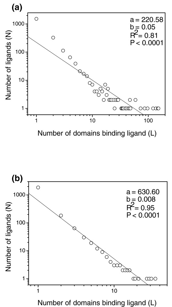
Results: By examining 2,186 well-defined small-molecule ligands and thousands of protein domains derived from a database of druggable binding sites, we show that a few ligands bind tens of protein domains or folds, whereas most ligands bind only one, which indicates that ligand-protein mapping follows a power law. Through assigning the protein-binding orders (early or late) for bio-ligands, we demonstrate that the preferential attachment principle still holds for the power-law relation between ligands and proteins. We also found that polar molecular surface area, H-bond acceptor counts, H-bond donor counts and partition coefficient are potential factors to discriminate ligands from ordinary molecules and to differentiate super ligands (shared by three or more folds) from others.
Conclusion: These findings have significant implications for evolution and drug discovery. First, the chronology of ligand-protein binding can be inferred by the power-law feature of ligand-protein mapping. Some nucleotide-containing ligands, such as ATP, ADP, GDP, NAD, FAD, dihydro-nicotinamide-adenine-dinucleotide phosphate (NDP), nicotinamide-adenine-dinucleotide phosphate (NAP), flavin mononucleotide (FMN) and AMP, are found to be the earliest cofactors bound to proteins, agreeing with the current understanding of evolutionary history. Second, the finding that about 30% of ligands are shared by two or more domains will help with drug discovery, such as in finding new functions from old drugs, developing promiscuous drugs and depending more on natural products.
Figures
Similar articles
-
Protein promiscuity: drug resistance and native functions--HIV-1 case.J Biomol Struct Dyn. 2005 Jun;22(6):615-24. doi: 10.1080/07391102.2005.10531228. J Biomol Struct Dyn. 2005. PMID: 15842167
-
Cofactor-binding sites in proteins of deviating sequence: comparative analysis and clustering in torsion angle, cavity, and fold space.Proteins. 2012 Feb;80(2):626-48. doi: 10.1002/prot.23226. Epub 2011 Nov 17. Proteins. 2012. PMID: 22095739
-
PDBLIG: classification of small molecular protein binding in the Protein Data Bank.J Med Chem. 2004 Jul 15;47(15):3807-16. doi: 10.1021/jm040804f. J Med Chem. 2004. PMID: 15239659
-
The structure of the protein universe and genome evolution.Nature. 2002 Nov 14;420(6912):218-23. doi: 10.1038/nature01256. Nature. 2002. PMID: 12432406 Review.
-
Implications of the small number of distinct ligand binding pockets in proteins for drug discovery, evolution and biochemical function.Bioorg Med Chem Lett. 2015 Mar 15;25(6):1163-70. doi: 10.1016/j.bmcl.2015.01.059. Epub 2015 Feb 3. Bioorg Med Chem Lett. 2015. PMID: 25690787 Free PMC article. Review.
Cited by
-
The phylogenomic roots of modern biochemistry: origins of proteins, cofactors and protein biosynthesis.J Mol Evol. 2012 Feb;74(1-2):1-34. doi: 10.1007/s00239-011-9480-1. Epub 2012 Jan 1. J Mol Evol. 2012. PMID: 22210458
-
Proteome evolution and the metabolic origins of translation and cellular life.J Mol Evol. 2011 Jan;72(1):14-33. doi: 10.1007/s00239-010-9400-9. Epub 2010 Nov 17. J Mol Evol. 2011. PMID: 21082171
-
Prebiotic Synthesis of ATP: A Terrestrial Volcanism-Dependent Pathway.Life (Basel). 2023 Mar 8;13(3):731. doi: 10.3390/life13030731. Life (Basel). 2023. PMID: 36983886 Free PMC article.
-
Phosfinder: a web server for the identification of phosphate-binding sites on protein structures.Nucleic Acids Res. 2011 Jul;39(Web Server issue):W278-82. doi: 10.1093/nar/gkr389. Epub 2011 May 26. Nucleic Acids Res. 2011. PMID: 21622655 Free PMC article.
-
Natural products and drug discovery. Can thousands of years of ancient medical knowledge lead us to new and powerful drug combinations in the fight against cancer and dementia?EMBO Rep. 2009 Mar;10(3):194-200. doi: 10.1038/embor.2009.12. Epub 2009 Feb 20. EMBO Rep. 2009. PMID: 19229284 Free PMC article. No abstract available.
References
Publication types
MeSH terms
Substances
LinkOut - more resources
Full Text Sources
Research Materials
