. 2007 Aug 29:4:61.
doi: 10.1186/1742-4690-4-61.

Targeting APOBEC3A to the viral nucleoprotein complex confers antiviral activity

Affiliations

Targeting APOBEC3A to the viral nucleoprotein complex confers antiviral activity

Ritu Goila-Gaur et al. Retrovirology. .

Abstract

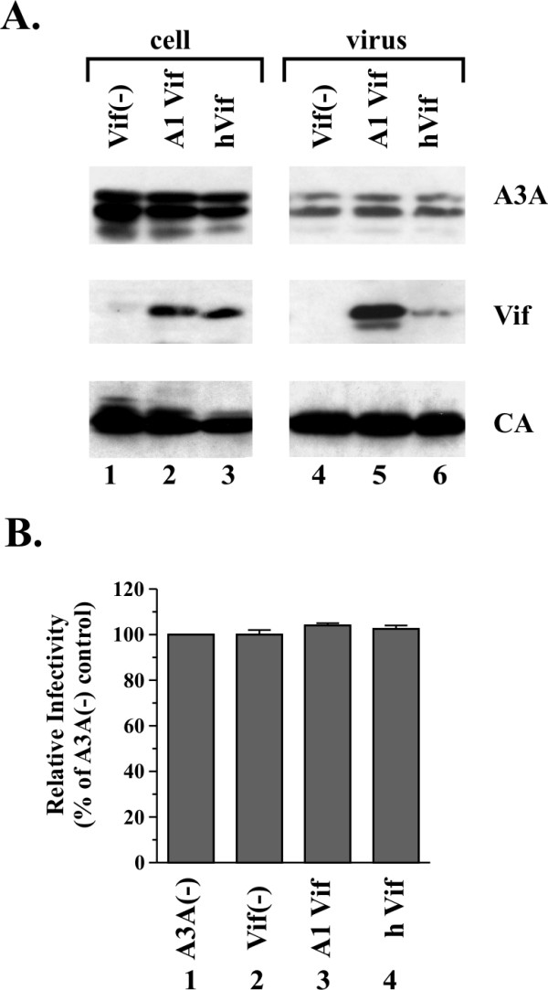
Background: APOBEC3 (A3) proteins constitute a family of cytidine deaminases that provide intracellular resistance to retrovirus replication and to transposition of endogenous retroelements. A3A has significant homology to the C-terminus of A3G but has only a single cytidine deaminase active site (CDA), unlike A3G, which has a second N-terminal CDA previously found to be important for Vif sensitivity and virus encapsidation. A3A is packaged into HIV-1 virions but, unlike A3G, does not have antiviral properties. Here, we investigated the reason for the lack of A3A antiviral activity.

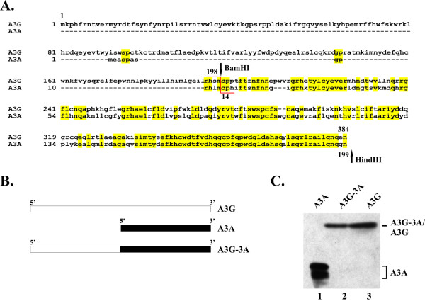
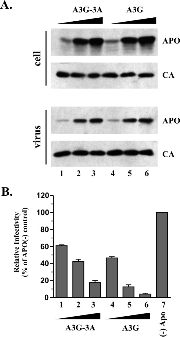
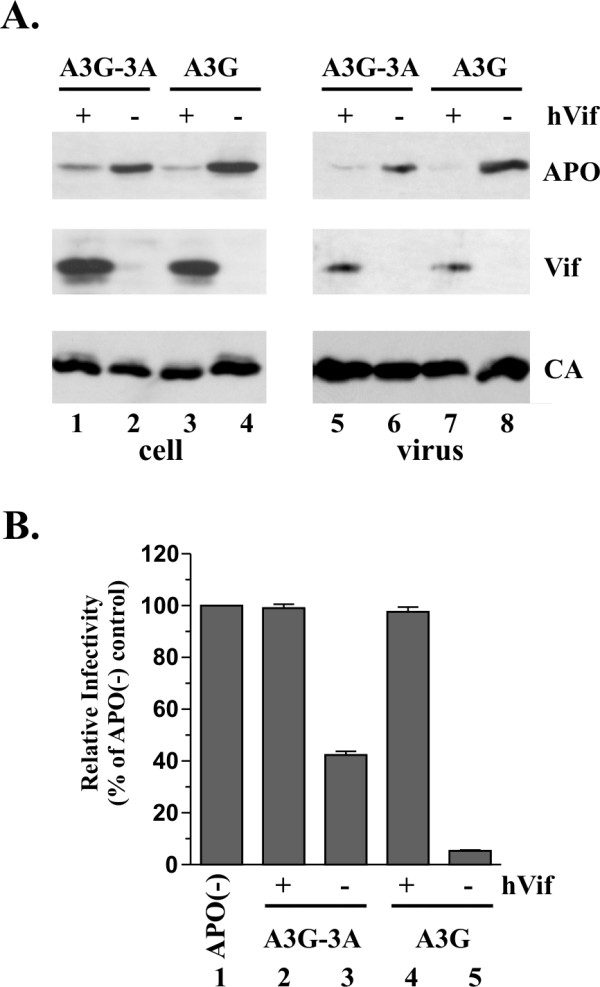
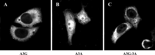
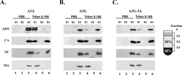
Results: Sequence alignment of A3G and A3A revealed significant homology of A3A to the C-terminal region of A3G. However, while A3G co-purified with detergent-resistant viral nucleoprotein complexes (NPC), virus-associated A3A was highly detergent-sensitive leading us to speculate that the ability to assemble into NPC may be a property conveyed by the A3G N-terminus. To test this model, we constructed an A3G-3A chimeric protein, in which the N-terminal half of A3G was fused to A3A. Interestingly, the A3G-3A chimera was packaged into HIV-1 particles and, unlike A3A, associated with the viral NPC. Furthermore, the A3G-3A chimera displayed strong antiviral activity against HIV-1 and was sensitive to inhibition by HIV-1 Vif.

Conclusion: Our results suggest that the A3G N-terminal domain carries determinants important for targeting the protein to viral NPCs. Transfer of this domain to A3A results in A3A targeting to viral NPCs and confers antiviral activity.


Figures

Figure 1
Figure 1
A3A is resistant to Vif induced degradation. (A) HeLa cells were transfected with vectors expressing vif-deficient pNL4-3 (3 μg each) along with pcDNA-A3A (1.5 μg each) and 1.5 μg of either pNL-A1vif(-) (lane 1), pNL-A1 (lane 2), or pcDNA-hVif (lane 3). Cells were harvested 24 h after transfection and whole-cell lysates were analyzed by immunoblotting using an A3G-specific rabbit polyclonal antibody (ApoC17) followed by incubation with an HRP-conjugated anti-rabbit antibody (A3A). The same blot was subsequently re-blotted with a Vif-specific monoclonal antibody (Vif) followed by probing with an HIV-positive patient serum to identify capsid protein (CA). Proteins are identified on the right. (B) Virus-containing supernatants from panel A were normalized for equivalent amounts of reverse transcriptase activity and used to infect LuSIV indicator cells [51] for determination of viral infectivity as described in Materials and Methods. Luciferase activity induced by virus produced in the absence of Vif and A3G was defined as 100% (lane 1). The infectivity of the remaining viruses was calculated relative to the control virus. Error bars reflect standard deviations from triplicate independent infections.
Figure 2
Figure 2
Construction and expression of A3G-3A chimera. (A) Sequence alignment of A3G and A3A. Highlighted areas indicate regions of amino acid identity. Arrows mark the location of unique BamHI and HindIII restriction sites in the expression vectors used for construction of the A3G-3A chimera. The chimera was constructed by replacing the BamHI and HindIII fragment in A3G by that of A3A. (B) Schematic illustration of the APOBEC expression vectors used in this study. (C) Expression of APOBEC proteins. HeLa cells were transfected with 5 μg each of pcDNA-A3A (lane 1), pcDNA-A3G-3A (lane 2), and pcDNA-A3G (lane 3). Total cell lysates were prepared 24 h after transfection and analyzed by immunoblotting for the expression of A3A, A3G-3A, and A3G, respectively using an A3G-specific polyclonal peptide antibody (ApoC17). Proteins are identified on the right.
Figure 3
Figure 3
The A3G-3A chimera has antiviral activity. (A) HeLa cells were transfected with vectors expressing vif-deficient pNL4-3 (3 μg each) along with increasing amounts of pcDNA-A3G-3A DNA (lane 1, 1 μg; lane 2, 2 μg; lane 3, 3 μg) or pcDNA-A3G DNA (lane 4, 0.2 μg; lane 5, 0.5 μg, lane 6, 1 μg). Higher amounts of A3G-3A DNA relative to A3G DNA were chosen because A3G-3A was generally expressed at lower levels than A3G. The total amount of transfected DNA in each sample was adjusted to 6 μg using empty pcDNA3.1 vector DNA. Cells and virus-containing supernatants were collected 24 h post-transfection. Total cell lysate and concentrated virus preparations were analyzed by immunoblotting using an A3G-specific rabbit polyclonal antibody (ApoC17) followed by incubation with an HRP-conjugated anti-rabbit antibody (APO). The same blot was subsequently re-blotted with an HIV-positive patient serum (CA). (B) Virus-containing supernatants from panel A were normalized for equal reverse transcriptase activity and used to infect LuSIV indicator cells [51] for determination of viral infectivity as described in Materials and Methods. Luciferase activity induced by virus produced in HeLa cells in the absence of Vif and A3G was defined as 100% infectivity (lane 7). The infectivity of the remaining viruses was calculated relative to the control virus. Error bars reflect standard deviations from triplicate independent infections.
Figure 4
Figure 4
A3G-3A is sensitive to HIV-1 Vif. (A) HeLa cells were transfected with vectors expressing vif-deficient pNL4-3 (3 μg each) along with 1.5 μg each of pcDNA-A3G-3A (lanes 1–2, 5–6) or pcDNA-Apo3G (lanes 3–4. 7–8) as well as 1.5 μg pcDNA-hVif (+) or 1.5 μg empty pcDNA3.1 vector DNA (-). Cells and virus-containing supernatants were collected 24 h post-transfection. Total cell lysates and concentrated virus preparations were analyzed by immunoblotting using an A3G-specific rabbit polyclonal antibody (ApoC17) followed by incubation with an HRP-conjugated anti-rabbit antibody (APO). The same blot was subsequently re-probed with a Vif-specific monoclonal antibody (Vif) followed by an HIV-positive patient serum (CA). Proteins are identified on the right. (B) Virus-containing supernatants from panel A were normalized for equal reverse transcriptase activity and used to infect LuSIV indicator cells to [51] determine viral infectivity as described in Materials and Methods. Luciferase activity induced by virus produced in HeLa cells in the absence of Vif and A3G was defined as 100% infectivity (lane 1). The infectivity of the remaining viruses was calculated relative to the control virus. Error bars reflect standard deviations from triplicate independent infections.
Figure 5
Figure 5
Subcellular localization of APOBEC proteins. HeLa cells were transfected with 5 μg each of pcDNA-Apo3G (panel A), pcDNA-A3A (panel B), or pcDNA-A3G-3A (panel C). Immediately following transfection, cells were detached from the flasks by trypsinization and re-seeded into 12 well plates containing microscope cover slips. Transfected cells were grown on the cover slips over night and then fixed with ice cold methanol for 10 minutes (-20°C). Cells were then stained with an A3G-specific rabbit polyclonal antibody (ApoC17) and analyzed by confocal microscopy as detailed in Materials and Methods.
Figure 6
Figure 6
A3G-3A co-purifies with viral nucleoprotein complexes. Virus stocks were made in HeLa cells by cotransfection of pNL4-3 Vif (-) plasmid DNA (3 μg each) with 2 μg each of pcDNA-A3A (panel A), pcDNA-A3G (panel B), or pcDNA-A3G-3A DNA (panel C). Virus containing supernatants were collected 24 h post-transfection, filtered to remove cellular debris, and concentrated by pelleting through 20% sucrose. Viral pellets were suspended in 1 ml of DMEM and 500 μl each of the virus preparation was loaded onto a 20%/60% sucrose step gradient previously overlaid with 100 μl of PBS (lanes 1–3) or Triton X-100 (lanes 4 to 6) as described in Materials and Methods. Three fractions of 1.1 ml each were collected from the top of the gradient as shown in the cartoon on the right. Fraction S1 (lanes 1 & 4) contains soluble proteins; fraction S2 (lanes 2 & 5) is a buffer fraction of 20% sucrose that separates soluble proteins from virus particles or viral cores; fraction S3 (lanes 3 & 6) includes the interphase of 20%:60% sucrose where viral particles and viral cores accumulate. Gradient fractions were subjected to immunoblot analysis using an A3G-specific antibody (A3A, A3G, or A3G-3A) followed by probing with an HIV-positive patient serum (CA). Nucleocapsid protein (NC) was identified by a goat anti-NC antibody and matrix protein (MA) was identified by a mouse monoclonal antibody to MA(P17).

Similar articles

Cited by

References

    1. Jarmuz A, Chester A, Bayliss J, Gisbourne J, Dunham I, Scott J, Navaratnam N. An anthropoid-specific locus of orphan C to U RNA-editing enzymes on chromosome 22. Genomics. 2002;79:285–296. doi: 10.1006/geno.2002.6718. - DOI - PubMed
    1. Wedekind JE, Dance GS, Sowden MP, Smith HC. Messenger RNA editing in mammals: new members of the APOBEC family seeking roles in the family business. Trends Genet. 2003;19:207–216. doi: 10.1016/S0168-9525(03)00054-4. - DOI - PubMed
    1. Sheehy AM, Gaddis NC, Choi JD, Malim MH. Isolation of a human gene that inhibits HIV-1 infection and is suppressed by the viral Vif protein. Nature. 2002;418:646–650. doi: 10.1038/nature00939. - DOI - PubMed
    1. Kao S, Khan MA, Miyagi E, Plishka R, Buckler-White A, Strebel K. The human immunodeficiency virus type 1 Vif protein reduces intracellular expression and inhibits packaging of APOBEC3G (CEM15), a cellular inhibitor of virus infectivity. J Virol. 2003;77:11398–11407. doi: 10.1128/JVI.77.21.11398-11407.2003. - DOI - PMC - PubMed
    1. Mangeat B, Turelli P, Caron G, Friedli M, Perrin L, Trono D. Broad antiretroviral defence by human APOBEC3G through lethal editing of nascent reverse transcripts. Nature. 2003;424:99–103. doi: 10.1038/nature01709. - DOI - PubMed

Publication types

MeSH terms