. 2007 Oct 1;310(1):140-53.
doi: 10.1016/j.ydbio.2007.07.038. Epub 2007 Aug 6.

TGF-beta mediated Msx2 expression controls occipital somites-derived caudal region of skull development

Affiliations

TGF-beta mediated Msx2 expression controls occipital somites-derived caudal region of skull development

Ryoichi Hosokawa et al. Dev Biol. .

Abstract

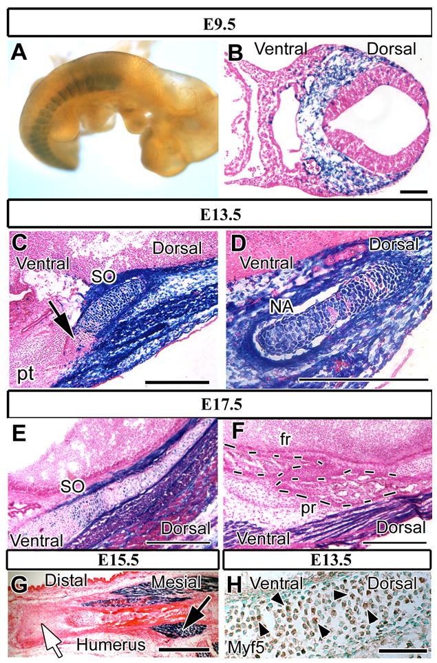
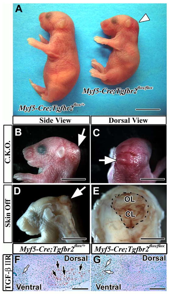
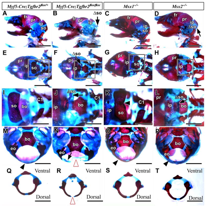
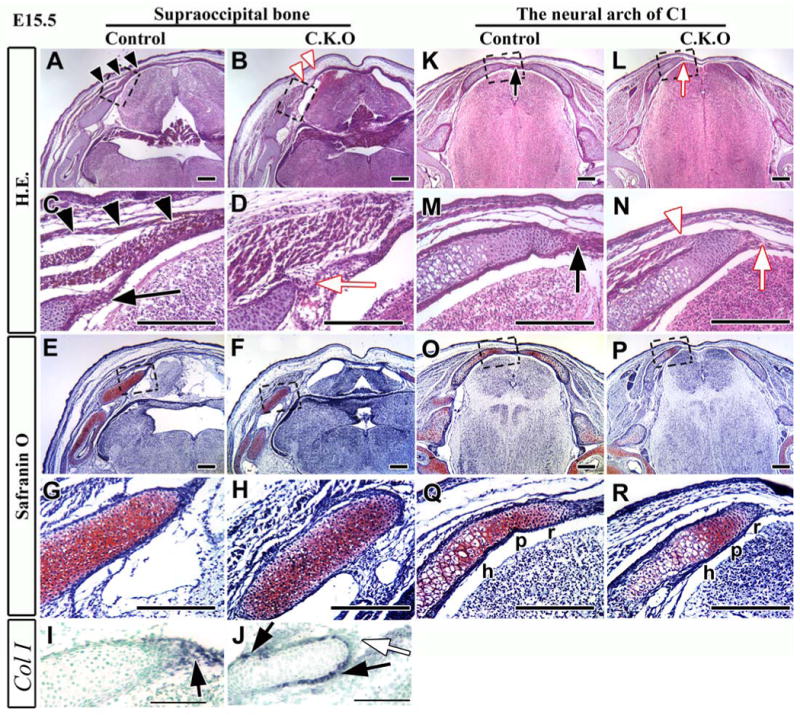
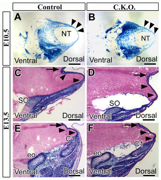
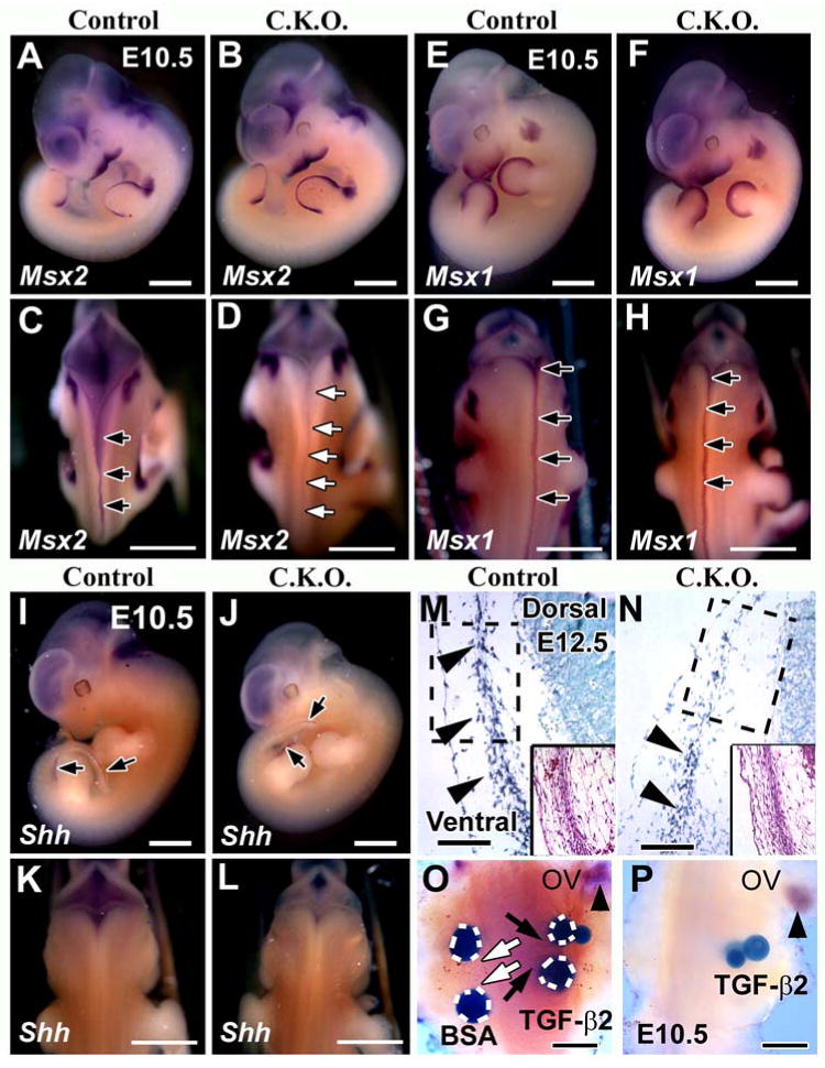
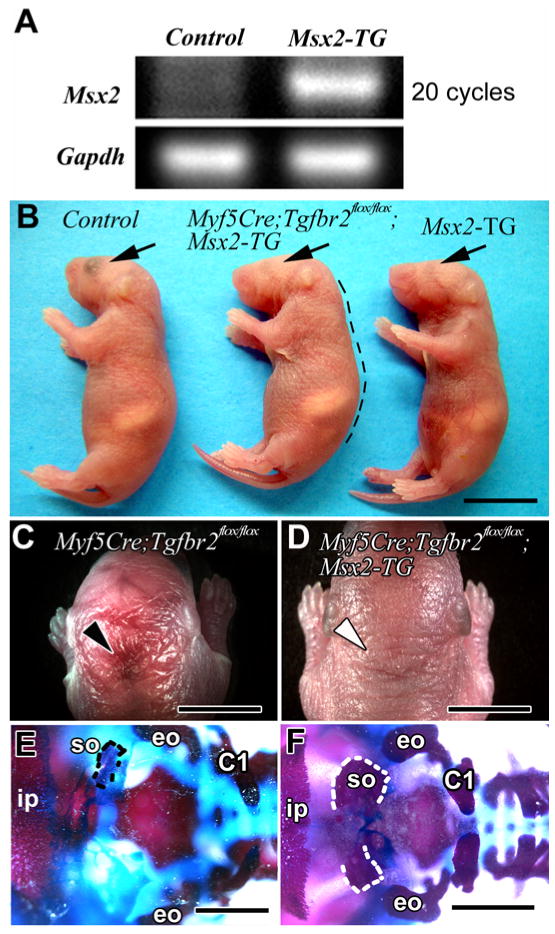
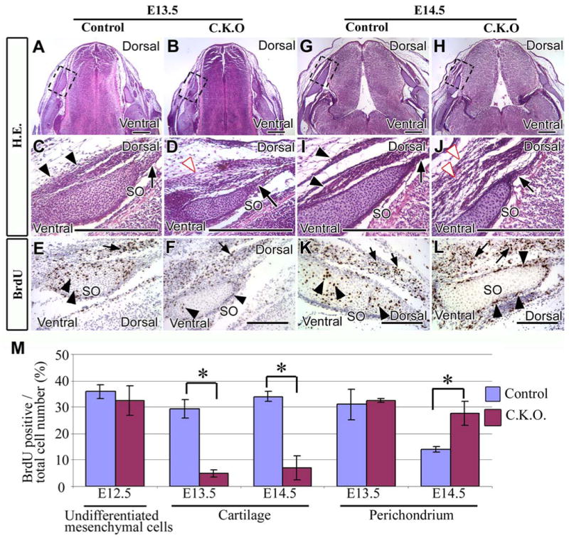
Craniofacial development involves cranial neural crest (CNC) and mesoderm-derived cells. TGF-beta signaling plays a critical role in instructing CNC cells to form the craniofacial skeleton. However, it is not known how TGF-beta signaling regulates the fate of mesoderm-derived cells during craniofacial development. In this study, we show that occipital somites contribute to the caudal region of mammalian skull development. Conditional inactivation of Tgfbr2 in mesoderm-derived cells results in defects of the supraoccipital bone with meningoencephalocele and discontinuity of the neural arch of the C1 vertebra. At the cellular level, loss of TGF-beta signaling causes decreased chondrocyte proliferation and premature differentiation of cartilage to bone. Expression of Msx2, a critical factor in the formation of the dorsoventral axis, is diminished in the Tgfbr2 mutant. Significantly, overexpression of Msx2 in Myf5-Cre;Tgfbr2flox/flox mice partially rescues supraoccipital bone development. These results suggest that the TGF-beta/Msx2 signaling cascade is critical for development of the caudal region of the skull.


Figures

Figure 1
Figure 1. Mesoderm-derived structures are positive for Myf5-directed expression of lacZ
Lateral view of lacZ staining (blue) of Myf5-Cre;R26Rflox/+ whole-mount E9.5 samples (A) and cross sections of E9.5 (B), E13.5 (C, D), E15.5 (G) and E17.5 samples (E, F). (A) LacZ staining is detectable in somites but not the paraxial mesoderm cells in the craniofacial region. (B) Mesoderm-derived cells in the occipital somite area show lacZ expression. (C, D) The primordia of the supraoccipital bone (SO) and the neural arch of C1 (C1) are populated by lacZ positive somite–derived cells. Non-blue cells are visible in the ventral area of the SO and the cartilage primordium of the petrous part of the temporal bone (pt) (C, black arrow). (E) Chondrocytes in the supraoccipital bone are lacZ positive. (F) Cells in the parietal bone (pr) and frontal bone (fr) (dashed line) are negative for lacZ staining. (G) Chondrocytes and osteoblasts in the humerus are lacZ negative (white arrow). Myogenic cells are LacZ positive around the humerus (black arrow). (H) Myf5 immunostaining of E13.5 Myf5-Cre;R26Rflox/+ mice. Chondrocytes in the supraoccipital bone primordium are positive for Myf5 (arrowheads). Scale bars: 100 μm in B, 500 μm in C-D, 300 μm in E-G, 50 μm in H.
Figure 2
Figure 2. Myf5-Cre;Tgfbr2flox/flox mouse pups show altered posture and meningoencephalocele in the occipital area
(A) Myf5-Cre;Tgfbr2flox/flox mice (right) display a hunchback phenotype and hemorrhage in the occipital area (white arrowhead). (B, C) Swelling is associated with the hemorrhage in the occipital area (white arrow) in the conditional knockout mouse (C.K.O.). (D, E) Peeling the skin above the swelling area reveals the excrescence of the brain in the occipital area (white arrows). (F, G) Immunostaining of TGF-β type II receptor in the supraoccipital bone primordium of Myf5-Cre;Tgfbr2flox/+ (F) and Myf5-Cre;Tgfbr2flox/flox (G) mice. Brown color indicates TGF-β type II receptor expression in the supraoccipital bone primordium (black arrows). White arrows indicate immunopositive cells in the lacZ negative area (see figure 1C). Abbreviations: CL, cerebellum; OL, occipital lobe. Scale bars: 1 cm in A, 5 mm in B-E, 100 μm in F, G.
Figure 3
Figure 3. Supraoccipital bone defect in Myf5-Cre;Tgfbr2flox/flox newborn pups
Alizarin red/Alcian blue-stained skeletons of Myf5-Cre;Tgfbr2flox/+ (control; A, E, I, M, Q), Myf5-Cre;Tgfbr2flox/flox (B, F, J, N, R), Msx1-/- (C, G, K, O, S), and Msx2-/- (D, H, L, P, T) mice, showing side (A-D), dorsal (E-L), and ventral (M-P) views and C1 vertebrae (Q-T). I, J, K, and L are enlarged from the boxes of E, F, G, and H, respectively. (A, E) The control mouse contains a frontal (fr), parietal (pr), interparietal (ip), and supraoccipital bone (so). (B, F, J, N) The supraoccipital bone of the conditional knockout mouse is hypomorphic (Λso, black arrow). (C, G, K) The Msx1-/- mouse shows normal development of the supraoccipital bone. (D, H, L) The Msx2-/- mouse shows a diminished supraoccipital bone (Λso, black arrow). (M) The occipital bone surrounding the foramen magnum dorsally (black arrowhead) is continuous in the control mouse. (N) The conditional knockout mouse shows a gap in the dorsal portion of occipital bone (white arrowhead) and a diminished supraoccipital bone (black arrow). (O, P) The Msx1-/- and Msx2-/- mice have continuous occipital bones forming the foramen magnum (black arrowheads). (Q, R) The conditional knockout mouse shows a disconnection dorsally in the neural arch of the C1 vertebra (white arrowhead). (S, T) The C1 vertebrae in both Msx1-/- and Msx2-/- mice appear normal. Abbreviations: bo, basioccipital bone; C1, the neural arch of first cervical vertebra; eo, exoccipital bone. Scale bars: 5 mm in A-H, 2 mm in I-T.
Figure 4
Figure 4. The supraoccipital bone and the C1 vertebra fail to fuse in the mid-dorsal area in Myf5-Cre;Tgfbr2flox/flox newborn mice
Hematoxylin and Eosin (H.E.) staining (A-D, M-P) and Safranin O staining (E-H, Q-T) of cross sections from the supraoccipital bone (A-L), and the neural arch of C1 vertebra (M-T) in control and Myf5-Cre;Tgfbr2flox/flox mice (C.K.O.). C, D, G, H, O, P, S, T are enlarged areas from the boxes in A, B, E, F, M, N, Q, and R, respectively. (A, C) The supraoccipital bone of control mice extends to the mid-dorsal area (black arrowheads) and contains cartilage structure at the base of the bone. (B, D) and Myf5-Cre;Tgfbr2flox/flox mice have a shorter extension of the supraoccipital bone (arrow indicates the edge of bone) and a conversion of cartilage into bone structure. Part of the brain is outside the skull vault (B, white arrow). (E, G) The cartilage in the supraoccipital bone base of control mice contains hypertrophic (h), proliferating (p), and resting (r) zones. (F, H) The base of the supraoccipital bone in the conditional knockout mice contains only a small hypertrophic zone, and the remainder is replaced by bone tissue (black arrow). (I, J) Type I collagen expression in the supraoccipital bone of control (I) and Myf5-Cre;Tgfbr2flox/flox (J) mice (black arrows). (K, L) Immunostaining of CD45, a marker of hematopoietic cells, in control (K) and Myf5-Cre;Tgfbr2flox/flox (L) mice. CD45 positive cells (brown color, arrowheads) are detectable adjacent to bone in the Myf5-Cre;Tgfbr2flox/flox sample. (M, O) The right and left sides of the neural arch of C1 vertebra are connected with cartilage mid-dorsally in the control mice. (N, P) The neural arch of C1 vertebra in Myf5-Cre;Tgfbr2flox/flox mice does not extend to the dorsal midline (asterisk). (Q, S) The mid-dorsal cartilage of control mice contains hypertrophic (h), proliferating (p), and resting (r) zones. (R, T) The edge of the neural arch, which does not extend to the midline, contains only a hypertrophic zone in the and Myf5-Cre;Tgfbr2flox/flox mice. Abbreviations: pt, petrous part of temporal bone; sc, spinal cord. Scale bars: 500 μm in A, B, E, F, M, N, Q, R, 300 μm in C, D, G, H, I, J, O, P, S, T, 50 μm in K, L.
Figure 5
Figure 5. Development of cartilage and muscle tissue is compromised in the mid-dorsal region of Myf5-Cre;Tgfbr2flox/flox mice
Hematoxylin and Eosin (A-D, K-N) and Safranin O (E-H, O-R) staining of cross-sections of the supraoccipital bone (A-H) and the C1 vertebra (K-R) in control and Myf5-Cre;Tgfbr2flox/flox mice. C, D, G, H, M, N, Q, and R are enlarged from the boxes of A, B, E, F, K, L, O, and P, respectively. (A, C) Muscle tissue forms continuous layers extending into the mid-dorsal region (black arrowheads) in control mice, and the dorsal tip of the perichondrium is expanding in the dorsal direction (black arrow). (B, D) Muscle tissue of conditional knockout mice does not extend into the mid-dorsal region (B, white arrowheads), and the perichondrium is disorganized at the dorsal tip (D, white arrow). (E-H) The primordium of the supraoccipital bone contains undifferentiated cartilage tissue in both control and conditional knockout mice. (I, J) Type I collagen (Col I) expression in control (I) and Myf5-Cre;Tgfbr2flox/flox (J) mice. Col I was expressed in the dorsal tip of the perichondrium (black arrow) in control mice. Col I expression was compromised in the dorsal tip (white arrow) of conditional knockout mice. Col I expression was detectable surrounding the membrane of the supraoccipital bone primordium (black arrows). (K, M) The perichondrium from both sides of the neural arch is fused in the mid-dorsal region in control mice (black arrow). (L, N) The neural arch has detached in the dorsal area of the conditional knockout mice (white arrow). Muscle has attached to bone at the tip of the neural arch of C1 vertebra (white arrowhead). (O-R) Both control and conditional knockout mice have three zones, hypertrophic (h), proliferative (p), and resting (r). Scale bars: 200 μm in A-H and K-R, 100 μm in I-J.
Figure 6
Figure 6. Cell migration from the dorsal sclerotome is unaffected in Myf5-Cre;Tgfbr2flox/flox mice
Cross sections stained for lacZ at E10.5 (A, B) and E13.5 (C-F). (A, B) Control (A: Myf5-Cre;Tgfbr2flox/+;R26Rflox/+) and conditional knockout (B: Myf5-Cre;Tgfbr2flox/flox;R26Rflox/+) mice contained mesoderm-derived cells (dark blue) migrating into the mid-dorsal area (black arrowheads). (C-F) Mesoderm-derived cells (black arrowheads) migrate to the mid-dorsal region in both control and conditional knockout mice, arriving at the supraoccipital bone (SO) and the neural arch of C1 vertebra (C1). Black arrows indicate the dorsal midline. Abbreviations: NT, neural tube; eo, exoccipital bone. Scale bars: 500 μm in A-F.
Figure 7
Figure 7. Msx2 gene expression is altered in the mid-dorsal area of Myf5-Cre;Tgfbr2flox/flox mice
Msx2 (A-D), Msx1 (E-H), and Shh (I-L) expression at E10.5 in control (A, C, E, G, I, K) and Myf5-Cre;Tgfbr2flox/flox mice (B, D, F, H, J, L). (A, B, E, F, I, J) lateral views. (C, D, G, H, K, L) dorsal views. (A, B) Msx2 expression is indistinguishable in lateral views of conditional knockout and control mice. (C, D) Msx2 is expressed in the mid-dorsal region extending from rostral to caudal (black arrows) in control mice. Msx2 expression is diminished in conditional knockout mice throughout the mid-dorsal region (white arrows). (E-H) Msx1 expression in conditional knockout mice is indistinguishable from control in the mid-dorsal line extending from rostral to caudal (black arrows). (I-L) Shh expression in conditional knockout mice is indistinguishable from the control in the hind limb and tail bud (I, J, black arrows). Shh was negative at the dorsal area in both the control and the conditional knockout mice (K, L). (M, N) Msx2 expression in mesenchyme cell cross sections at E12.5. Inserts show H.E. staining. Msx2 expression is visible throughout the ventral region towards the dorsal region in the control mice (M, black arrowheads). Msx2 expression is restricted to the ventral side of conditional knockout mice (N, black arrowheads). (O, P) Explants from the mid-dorsal area of E10.5 control and conditional knockout mice were treated with beads for 12 hours. The control sample (O) treated with TGF-β2 beads is positive for Msx2 expression (black arrows, dark blue), but BSA beads do not induce Msx2 expression (white arrows). The conditional knockout sample treated with TGF-β2 beads is negative for Msx2 expression (P). Msx2 gene expression is visible around the otic vesicle (ov) in both samples (black arrowhead). Scale bars: 1 mm in A-L, 100 μm in M, N, 200 μm in O, P.
Figure 8
Figure 8. Msx2 overexpression rescues the supraoccipital bone defect in Myf5-Cre;Tgfbr2flox/flox mice
(A) Msx2 gene expression detected by PCR in control (Tgfbr2flox/+) and Msx2-TG (Msx2Tg/+;Tgfbr2flox/+) samples at E10.5. (B) Whole mount views of Myf5-Cre;Tgfbr2flox/+ (control), Myf5-Cre;Tgfbr2flox/flox;Msx2-TG (transgenic) and Msx2-TG (transgenic) mice. The Myf5-Cre;Tgfbr2flox/flox;Msx2-TG mouse has a hunchback phenotype like Myf5-Cre;Tgfbr2flox/flox mice (black dotted line) and microphthalmia like the Msx2-TG mouse (black arrows). (C) Dorsal view of the Myf5-Cre;Tgfbr2flox/flox mouse shows the hemorrhage around the occipital area (black arrowhead). (D) Dorsal view of the Myf5-Cre;Tgfbr2flox/flox;Msx2-TG mouse shows no hemorrhage around the occipital area (white arrowhead). (E, F) Alizarin red and alcian blue staining. In a dorsal view, the Myf5-Cre;Tgfbr2flox/flox mouse has a diminished supraoccipital bone (E, black dotted line). The base of the supraoccipital bone is formed but each side is disconnected in the mid-dorsal area (F, white dotted line) in the Myf5-Cre;Tgfbr2flox/flox;Msx2-TG mouse. Abbreviations: C1, the neural arch of C1 vertebra; eo, exoccipital bone; ip, interparietal bone. Scale bars: 1 cm in B, 5 mm in C, D, 2 mm in E, F.
Figure 9
Figure 9. The pattern of cell proliferation is altered in the supraoccipital bone primordium of Myf5-Cre;Tgfbr2flox/flox mice
Cross sections of E13.5 (A-F) or E14.5 (G-L) supraoccipital bone primordium stained with hematoxylin and eosin (A-D, G-J) or BrdU (E, F, K, L). (C-F, I-L) Enlarged areas from the dashed black boxes of A, B, G, and H are shown in C/E, D/F, I/K, and J/L, respectively. (A, C) Control mice have well-organized perichondrium (C, black arrow) and muscle tissue (C, black arrowheads). (B, D) The perichondrium of conditional knockout mice is organized (D, black arrow), but muscle tissue is disorganized (D, white arrowhead). (E, F) Control and conditional knockout mice contain BrdU positive cells in the cartilage and the perichondrium of the supraoccipital bone (black arrowheads) and in the muscle cells (black arrow). (G, I) The perichondrium (I, black arrow) and muscle tissue (I, black arrowheads) are well-organized in control mice. (H, J) Conditional knockout mice have a well-organized perichondrium (black arrow), but disorganized muscle tissue (J, white arrowheads). (K, L) In control mice, BrdU positive cells are present in both the cartilage and perichondrium of the supraoccipital bone (black arrowheads) and also in the muscle (black arrows). BrdU positive cells are visible in the muscle (black arrows) and perichondrium (black arrowheads) of the supraoccipital bone in conditional knockout mice, but not in the cartilage of supraoccipital bone. (M) Statistical analysis of cell proliferation activity in control and conditional knockout mice. Five randomly selected, non-overlapping samples were used to obtain the BrdU labeling index from each experimental group. Student t-tests were used for statistical analysis. *:P<0.05. Abbreviations: SO, the primordium supraoccipital bone. Scale bars: 300 μm in A-L.

Similar articles

Cited by

References

    1. Alappat S, Zhang ZY, Chen YP. Msx homeobox gene family and craniofacial development. Cell Res. 2003;13:429–442. - PubMed
    1. Alvarez J, Horton J, Sohn P, Serra R. The perichondrium plays an important role in mediating the effects of TGF-beta1 on endochondral bone formation. Dev Dyn. 2001;221:311–321. - PubMed
    1. Alvarez J, Sohn P, Zeng X, Doetschman T, Robbins DJ, Serra R. TGFbeta2 mediates the effects of hedgehog on hypertrophic differentiation and PTHrP expression. Development. 2002;129:1913–1924. - PubMed
    1. Bach A, Lallemand Y, Nicola MA, Ramos C, Mathis L, Maufras M, Robert B. Msx1 is required for dorsal diencephalon patterning. Development. 2003;130:4025–4036. - PubMed
    1. Baffi MO, Slattery E, Sohn P, Moses HL, Chytil A, Serra R. Conditional deletion of the TGF-beta type II receptor in Col2a expressing cells results in defects in the axial skeleton without alterations in chondrocyte differentiation or embryonic development of long bones. Dev Biol. 2004;276:124–142. - PubMed

Publication types

LinkOut - more resources