. 2007 Dec 5;369(1):12-8.
doi: 10.1016/j.virol.2007.07.023. Epub 2007 Aug 28.

Stable cell lines expressing high levels of the herpes simplex virus type 1 LAT are refractory to caspase 3 activation and DNA laddering following cold shock induced apoptosis

Affiliations

Stable cell lines expressing high levels of the herpes simplex virus type 1 LAT are refractory to caspase 3 activation and DNA laddering following cold shock induced apoptosis

Dale Carpenter et al. Virology. .

Abstract

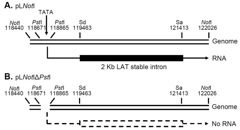
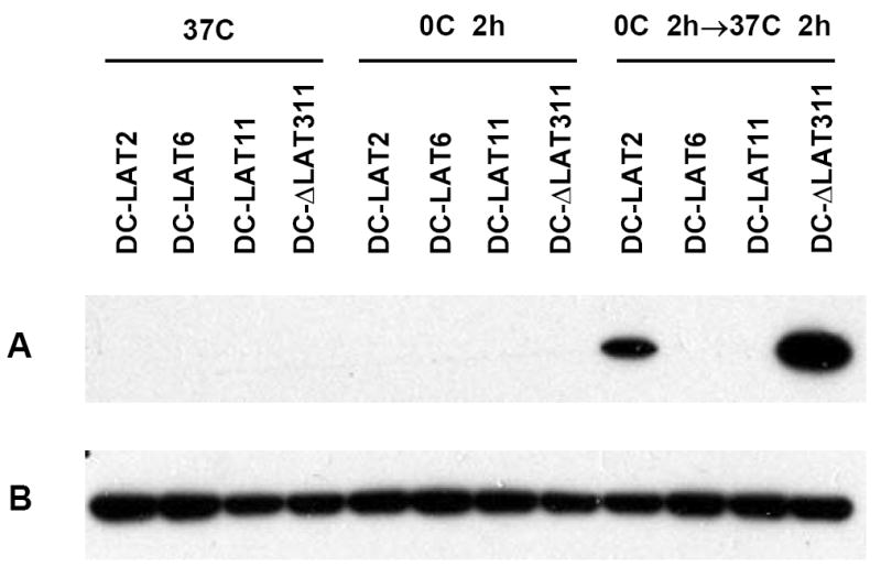
The herpes simplex virus type 1 (HSV-1) latency associated transcript (LAT) gene's anti-apoptosis activity plays a central, but not fully elucidated, role in enhancing the virus's reactivation phenotype. In transient transfection experiments, LAT increases cell survival following an apoptotic insult in the absence of other HSV-1 genes. However, the high background of untransfected cells has made it difficult to demonstrate that LAT inhibits specific apoptotic factors such as caspases. Here we report that, in mouse neuroblastoma cell lines (C1300) stably expressing high levels of LAT, cold shock induced apoptosis was blocked as judged by increased survival, protection against DNA fragmentation (by DNA ladder assay), and inhibition of caspase 3 cleavage and activation (Western blots). To our knowledge, this is the first report providing direct evidence that LAT blocks two biochemical hallmarks of apoptosis, caspase 3 cleavage and DNA laddering, in the absence of other HSV-1 gene products.


Figures

Figure 1
Figure 1
Schematic diagram of the cloned LAT region expressed in stable C1300 cell lines. Panel A: pLNotI, a 3,586bp NotI-NotI DNA restriction fragment containing LAT promoter nts -361 to +1 followed by the first 3225 nts of the primary LAT transcript (+1 to +3225). This restriction fragment was used to generate the cell lines DC-LAT2, DC-LAT6, and DC-LAT11. The LAT RNA expressed is shown under the plasmid. Panel B: pLNotPstI is identical to pLNotI but with a PstI-PstI deletion corresponding to LAT nts -130 to +64. This deletion removes essential LAT promoter elements including the LAT TATA box and no detectable LAT RNA is made (dashed lines). This plasmid was used to generate DC-ΔLAT35 and DC-ΔLAT311 cells. Sd=Splice donor; Sa=Spice acceptor.
Figure 2
Figure 2
Northern blot of LAT-RNA in stable C1300 derived cell lines. Total RNA was isolated from cells and analyzed by Northern blot using a probe specific for the stable 2 kb LAT intron as described in Materials and Methods. The cell lines are indicated at the top. The arrow indicates the location of 18S ribosomal RNA (with an apparent mobility of 2 kb) based on ethidium bromide staining.
Figure 3
Figure 3
Cell survival following cold shock. Each cell line was plated into 6 T25 flasks and allowed to recover overnight in a 37 °C CO2 incubator. The next day the flasks were tightly capped. Three flasks of each cell line were incubated for 2 hrs on ice (cold shocked) and 3 flasks of each cell line were incubated at 37 °C without CO2 (not cold shocked). After 2 hrs the caps were loosened and all flasks were returned to a 37 °C CO2 incubator to recover for 4 hours. After treatment the cells were washed once, trypsinized, and the number of cells that had remained attached was determined using a hemacytometer. The bars represent the average of 3 repeats +/- SD. The numbers above each bar indicate the percent of cells after cold shock [(cold shock/no cold shock) × 100%] for each cell line. The results for each cell line are representative of at least 3 independent experiments.
Figure 4
Figure 4
Decreased DNA laddering in LAT(+) cell lines following cold shock. Cells were maintained at 37 °C, cold shocked for 2 hours but not recovered, or cold shocked for 2 hours and recovered at 37 °C for 2hours as indicated. Fragmented chromosomal DNA was isolated using a hypotonic buffer that releases fragmented but not intact chromosomal DNA from the nucleus and equal aliquots were run on a gel as described in Materials and Methods.
Figure 5
Figure 5
LAT(+) cell lines are protected against cold shock induced caspase 3 cleavage. Cells were maintained at 37 °C (left panel) cold shocked and harvested immediately (middle panel) or allowed to recover for 2 hours at 37 °C (right panel) as described in Materials and Methods. Western blotting was done using an antibody specific for activated (cleaved) caspase 3 as described in Materials and Methods. A. Cleaved caspase-3. B. GAPDH loading controls.

References

    1. Ahmed M, Lock M, Miller CG, Fraser NW. Regions of the herpes simplex virus type 1 latency-associated transcript that protect cells from apoptosis in vitro and protect neuronal cells in vivo. J Virol. 2002;76(2):717–29. - PMC - PubMed
    1. Atanasiu D, Kent JR, Gartner JJ, Fraser NW. The stable 2-kb LAT intron of herpes simplex stimulates the expression of heat shock proteins and protects cells from stress. Virology. 2006;350(1):26–33. - PubMed
    1. Aubert M, Blaho JA. The herpes simplex virus type 1 regulatory protein ICP27 is required for the prevention of apoptosis in infected human cells. J Virol. 1999;73(4):2803–13. - PMC - PubMed
    1. Block TM, Deshmane S, Masonis J, Maggioncalda J, Valyi-Nagi T, Fraser NW. An HSV LAT null mutant reactivates slowly from latent infection and makes small plaques on CV-1 monolayers. Virology. 1993;192(2):618–630. - PubMed
    1. Bloom DC, Devi-Rao GB, Hill JM, Stevens JG, Wagner EK. Molecular analysis of herpes simplex virus type 1 during epinephrine- induced reactivation of latently infected rabbits in vivo. J Virol. 1994;68(3):1283–1292. - PMC - PubMed

Publication types