. 2007 Nov;81(21):12019-28.
doi: 10.1128/JVI.00300-07. Epub 2007 Aug 29.

Characterization of the early events in dengue virus cell entry by biochemical assays and single-virus tracking

Affiliations

Characterization of the early events in dengue virus cell entry by biochemical assays and single-virus tracking

Hilde M van der Schaar et al. J Virol. 2007 Nov.

Abstract

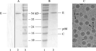
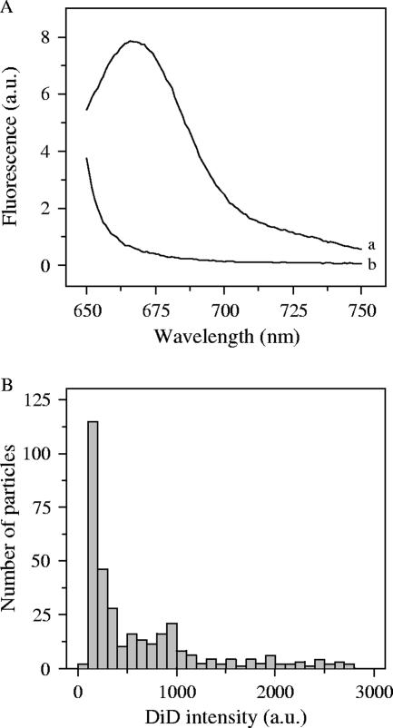
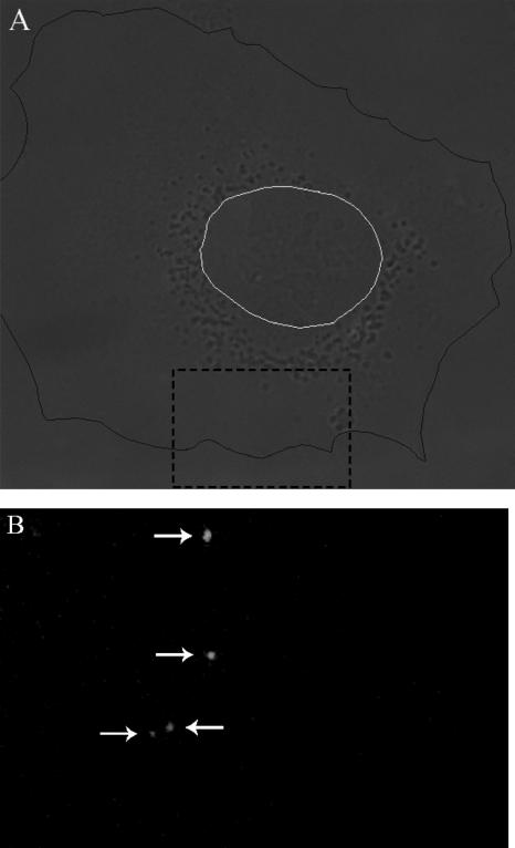
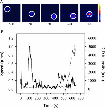
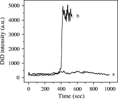
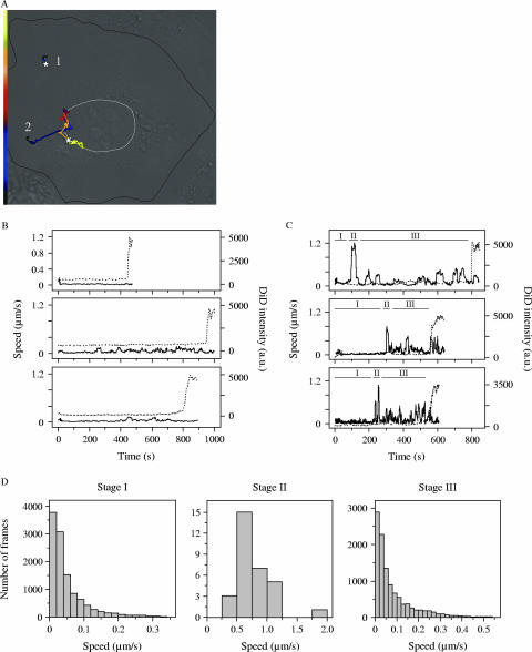
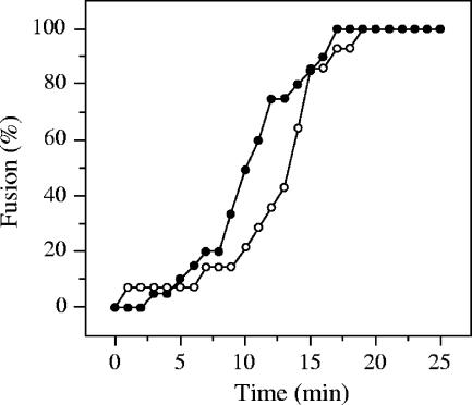
In this study, we investigated the cell entry characteristics of dengue virus (DENV) type 2 strain S1 on mosquito, BHK-15, and BS-C-1 cells. The concentration of virus particles measured by biochemical assays was found to be substantially higher than the number of infectious particles determined by infectivity assays, leading to an infectious unit-to-particle ratio of approximately 1:2,600 to 1:72,000, depending on the specific assays used. In order to explain this high ratio, we investigated the receptor binding and membrane fusion characteristics of single DENV particles in living cells using real-time fluorescence microscopy. For this purpose, DENV was labeled with the lipophilic fluorescent probe DiD (1,1'-dioctadecyl-3,3,3',3'-tetramethylindodicarbocyanine, 4-chlorobenzenesulfonate salt). The surface density of the DiD dye in the viral membrane was sufficiently high to largely quench the fluorescence intensity but still allowed clear detection of single virus particles. Fusion of the viral membrane with the cell membrane was evident as fluorescence dequenching. It was observed that DENV binds very inefficiently to the cells used, explaining at least in part the high infectious unit-to-particle ratio. The particles that did bind to the cells showed different types of transport behavior leading to membrane fusion in both the periphery and perinuclear regions of the cell. Membrane fusion was observed in 1 out of 6 bound virus particles, indicating that a substantial fraction of the virus has the capacity to fuse. DiD dequenching was completely inhibited by ammonium chloride, demonstrating that fusion occurs exclusively from within acidic endosomes.


Figures

FIG. 1.
FIG. 1.
Analysis of purified DENV grown on C6/36 cells. (A) Western blot analysis using MAB8702 antibody against DENV E glycoprotein. Lane 1, mock-infected cells; lane 2, purified DENV; lane 3, protein marker. (B) Gel stained with Coomassie blue. Lane 1, protein marker; lane 2, purified DENV. The positions of the bands for proteins E (55 kDa), prM (18.44 kDa), and C (12.55 kDa) and of the marker are indicated. The viral M protein (8.3 kDa) could not be detected, presumably due to its small size. (C) Cryoelectron micrograph of purified DENV S1. Immature (“spikey”) particles (1) and mature (“smooth”) particles (2) are indicated.
FIG. 2.
FIG. 2.
Characterization of DiD-labeled DENV particles. (A) Fluorescence emission spectra of DiD-labeled DENV. DiD-labeled DENV was mixed with HNE buffer at 37°C. Emission scans were recorded at wavelengths 650 to 750 nm with excitation at 640 ± 4 nm. Curve a, DiD-labeled DENV in presence of C12E8; curve b, DiD-labeled DENV. (B) The intensity of DiD-labeled DENV was determined with fluorescence microscopy and plotted in a histogram, as described in Materials and Methods. Particles with low fluorescence intensity (0 to 500 AU) were selected for image analysis.
FIG. 3.
FIG. 3.
Binding of DiD-labeled DENV to cells. Virus binding was determined by fluorescence microscopy as described in Materials and Methods. (A) Cell image obtained with DIC optics. The plasma membrane is indicated in black, and the nucleus is in white. (B) Fluorescent image of a small portion of the cell shown in panel A (dashed black square) to clearly demonstrate the virus spots. In this image, four virus spots can be seen.
FIG. 4.
FIG. 4.
Infectious properties of unbound DENV particles in cells. Virus binding was allowed for 15 min at 4°C on BS-C-1 cells, after which the unbound virus particles were collected and transferred to new cells. This procedure was repeated 10 times. Infection was analyzed by E protein expression in cells, and the titer was calculated as described in Material and Methods.
FIG. 5.
FIG. 5.
Trajectory of a single DENV particle in a cell. (A) Fluorescence images show one DiD-labeled DENV particle (surrounded by a white circle) at 560, 580, 600, 620, and 640 s after the temperature shift to 37°C. The colored bar indicates the DiD fluorescence intensity from low (black) to high (yellow) intensity. (B) Time trajectories of DiD fluorescence intensity (dashed line) and velocity (solid line) of the same virus particle. Membrane fusion of the virus particle with the endosomal membrane is detected as an increase in DiD fluorescence intensity. The arrow indicates the time point of membrane fusion. a.u., arbitrary units.
FIG. 6.
FIG. 6.
DENV entry in cells (ammonium chloride treated). Tracking analysis was performed as described in Materials and Methods. Curve a, time trajectory of the fluorescence intensity of DiD-labeled DENV in ammonium chloride-treated cells; curve b, untreated cells.
FIG. 7.
FIG. 7.
Distinct transport behavior of DENV particles in cells. (A) Two virus trajectories in a cell. The morphology of the cell was visualized with DIC optics as described in the legend of Fig. 3. The trajectories are color coded for time to indicate the velocity of the virus particle. The white stars represent the fusion sites. Trajectory 1, virus transport in the cell periphery with time axis from 0 s (black) to 450 s (yellow); trajectory 2, long-range three-stage movement toward the nucleus with time axis from 0 s (black) to 800 s (yellow). (B and C) Three time trajectories of DiD fluorescence intensity (dashed line) and speed (solid line) of stationary and long-range three-stage transport behavior, respectively. The upper graphs in both panels are trajectories 1 and 2 shown in panel A, respectively. In panel C the stages in the long-range three-stage movement are indicated (I, II, and III). (D) Quantitative analysis of the velocity in stages I, II, and III. For every frame within each stage, the velocities of all 30 virus particles that showed long-range three-stage movement are plotted.
FIG. 8.
FIG. 8.
Kinetics of DENV fusion in cells. The time point of membrane fusion was defined as described in the legend of Fig. 5. In total, 14 membrane fusion events in the cell periphery (open circles) were detected, and 30 were detected in the perinuclear region (closed circles). Time point 0 min is the moment of the temperature shift to 37°C. The fraction of fused virus particles was plotted as a function of time.

References

    1. Allison, S. L., J. Schalich, K. Stiasny, C. W. Mandl, and F. X. Heinz. 2001. Mutational evidence for an internal fusion peptide in flavivirus envelope protein E. J. Virol. 75:4268-4275. - PMC - PubMed
    1. Allison, S. L., J. Schalich, K. Stiasny, C. W. Mandl, C. Kunz, and F. X. Heinz. 1995. Oligomeric rearrangement of tick-borne encephalitis virus envelope proteins induced by an acidic pH. J. Virol. 69:695-700. - PMC - PubMed
    1. Anderson, R., S. Wang, C. Osiowy, and A. C. Issekutz. 1997. Activation of endothelial cells via antibody-enhanced dengue virus infection of peripheral blood monocytes. J. Virol. 71:4226-4232. - PMC - PubMed
    1. Bae, H. G., A. Nitsche, A. Teichmann, S. S. Biel, and M. Niedrig. 2003. Detection of yellow fever virus: a comparison of quantitative real-time PCR and plaque assay. J. Virol. Methods 110:185-191. - PubMed
    1. Chen, Y., T. Maguire, R. E. Hileman, J. R. Fromm, J. D. Esko, R. J. Linhardt, and R. M. Marks. 1997. Dengue virus infectivity depends on envelope protein binding to target cell heparan sulfate. Nat. Med. 3:866-871. - PubMed

Publication types

Substances