. 2007 Nov;81(22):12582-95.
doi: 10.1128/JVI.00381-07. Epub 2007 Aug 29.

Predominant mode of human immunodeficiency virus transfer between T cells is mediated by sustained Env-dependent neutralization-resistant virological synapses

Affiliations

Predominant mode of human immunodeficiency virus transfer between T cells is mediated by sustained Env-dependent neutralization-resistant virological synapses

Ping Chen et al. J Virol. 2007 Nov.

Abstract

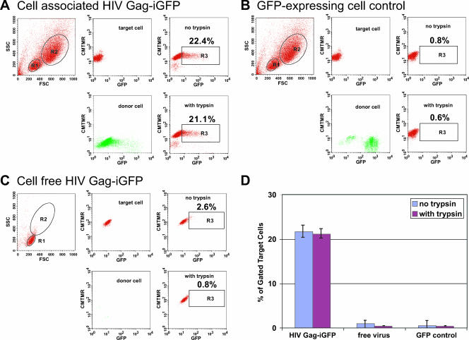
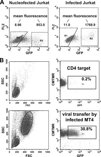
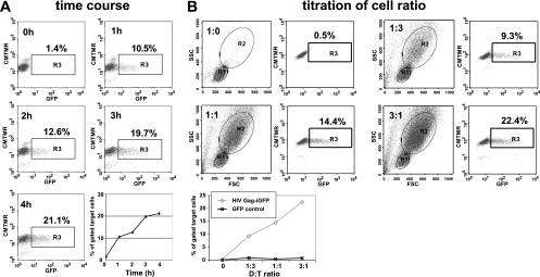
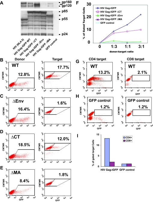
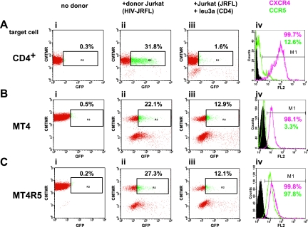
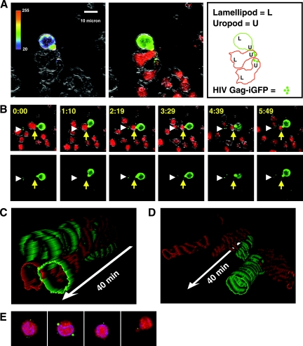
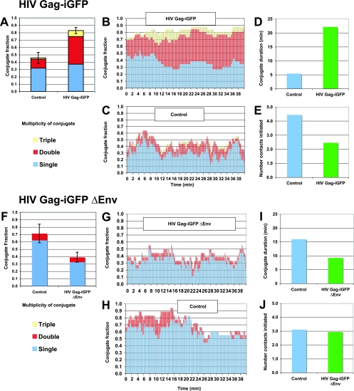
Cell-free human immunodeficiency virus type 1 (HIV-1) can initiate infections, but contact between infected and uninfected T cells can enhance viral spread through intercellular structures called virological synapses (VS). The relative contribution of VS to cell-free viral transfer has not been carefully measured. Using an ultrasensitive, fluorescent virus transfer assay, we estimate that when VS between HIV-expressing Jurkat T cells and primary CD4(+) T cells are formed, cell-associated transfer of virus is 18,000-fold more efficient than uptake of cell-free virus. Furthermore, in contrast to cell-free virus uptake, the VS deposits virus rapidly into focal, trypsin-resistant compartments in target T cells. This massive virus internalization requires Env-CD4 receptor interactions but is resistant to inhibition by patient-derived neutralizing antisera that inhibit homologous cell-free virus. Deleting the Env cytoplasmic tail does not abrogate VS-mediated transfer, but it renders the VS sensitive to neutralizing antibodies, suggesting that the tail limits exposure of VS-neutralizing epitopes on the surface of infected cells. Dynamic live imaging of the VS reveals that HIV-expressing cells are polarized and make sustained, Env-dependent contacts with target cells through uropod-like structures. The polarized T-cell morphology, Env-CD4 coordinated adhesion, and viral transfer from HIV-infected to uninfected cells suggest that VS allows HIV-1 to evade antibody neutralization and to disseminate efficiently. Future studies will discern to what extent this massive viral transfer contributes to productive infection or viral dissemination through the migration of virus-carrying T cells.


Figures

FIG. 1.
FIG. 1.
Massive VS-mediated transfer of HIV to target cells measured by flow-cytometry-based detection of HIV Gag-iGFP. (A) Target cells, which were CMTMR-labeled CD4+ primary T cells (R1 gate), were mixed with donor cells, which were HIV Gag-iGFP-expressing Jurkat cells (R2 gate), resulting in transfer of fluorescent virus into target cells (upper right). Middle panels display target and donor cell fluorescence prior to mixing. Trypsin-treated cells (lower right) did not decrease the signal. (B) Target cells mixed with control GFP-expressing cells did not show any viral transfer. (C) Target cells were mixed with 100 ng/ml of cell-free HIV Gag-iGFP fluorescent virus, resulting in weak fluorescence in a small percentage of cells. Numbers within the R3 gate indicate the percentage of target cells acquiring strong GFP fluorescence. (D) The average percentages of target cells acquiring GFP fluorescence were plotted from values obtained from at least three independent experiments with different donor cells, performed as described for panels A, B, and C. FSC, forward scatter; SSC, side scatter.
FIG. 2.
FIG. 2.
HIV Gag-iGFP-infected cells can engage in VS-mediated transfer. (A) Levels of HIV Gag-iGFP expressed by Amaxa-nucleofected Jurkat cells are comparable to those of HIV Gag-iGFP-infected Jurkat cells at 48 h after nucleofection/infection. Infection of Jurkat cells employed a modified spinoculation method (46) to achieve maximal efficiency. (B) HIV Gag-iGFP-infected MT4 cells can function as donor cells in VS-mediated viral transfer into primary CD4+ T cells. Target cells alone (top panels) or target cells plus infected MT4 cells (bottom panels) are shown. FSC, forward scatter; SSC, side scatter; FL2, fluorescence channel 2.
FIG. 3.
FIG. 3.
VS-mediated viral transfer is rapid and dependent on donor target ratio. (A) A 1:1 ratio of HIV Gag-iGFP-expressing donor cells and CD4+ target cells was mixed and incubated for 0, 1, 2, 3, or 4 h. The percentage of GFP+ cells is plotted over time (lower right). (B) Cells were mixed at donor/target ratios of 0:1, 1:3, 1:1, or 3:1 and were assayed after 3 h of transfer. The percentage of GFP+ cells following exposure to HIV Gag-iGFP-expressing cells is compared to that after exposure to control cells expressing GFP alone (bottom panel).
FIG. 4.
FIG. 4.
Efficient VS-mediated viral transfer is dependent upon Env and Gag. (A) Western blot of cell lysates of viral mutants nucleofected into Jurkat T cells and probed with anti-Env antibodies (top) or anti-HIV antisera (bottom). (B) HIV Gag-iGFP fluorescence in the donor cells (left) and in the target cells (right) after 3 h of coincubation. (C) Viral transfer assay on the Env-deleted clone ΔEnv. (D) Viral transfer assay on the cytoplasmic-tail-deleted clone ΔCT. (E) Viral transfer assay on the globular head of the MA deletion construct ΔMA. (F) Titration of the donor/target ratios of the various mutant clones. (G) CD8+ T cells do not engage in viral transfer with HIV Gag-iGFP-expressing donor cells. (H) Incubation of CD4+ or CD8+ cells with control GFP-expressing cells. (I) Comparison of CD4 to CD8 cells as target cells, graphing the percentage of GFP+ cells from experiments depicted in G and H. WT, wild type.
FIG. 5.
FIG. 5.
Viral transfer mediated through R5-tropic Env from molecular clone JRFL occurs efficiently in CCR5+ or CCR5 CD4+ T cells. (A) VS-mediated transfer from HIV Gag-iGFP(JRFL)-expressing Jurkat cells into primary CD4+ T cells shows efficient transfer into target cells. A parallel analysis of coreceptor expression in the target cells shows a small fraction of CCR5+ T cells. (B) VS-mediated transfer into MT4 does not require CCR5. (C) VS-mediated transfer into CCR5-expressing MT4R5 cells is similar to that for CCR5 MT4. Panel i, target cells alone; panel ii, target cells plus HIV Gag-iGFP(JRFL)-expressing Jurkat donor cells; panel iii, donor and target cells inhibited with 0.5 μg/ml of the anti-CD4 blocking antibody Leu 3a; panel iv, chemokine receptor staining of the target cells, illustrating the percentage of cells expressing CXCR4 (red) or CCR5 (green). FL2, fluorescence channel 2.
FIG. 6.
FIG. 6.
Live imaging of VS formation reveals polarized HIV-infected and uninfected cells engaging in long-lived contacts. (A) An HIV Gag-iGFP-expressing donor cell bound to a target cell through uropod-like structures. (Left) At the VS, increased concentrations of Gag-iGFP in the donor cell at the site of cell-cell contact were observed, with GFP fluorescence in a rainbow intensity scale overlaid on a DIC edge image to show the cell boundaries. (Middle) Three-color image with Gag-iGFP (green), a CMTMR-labeled target cell (red), and DIC edges (gray). (Right) Outlines of a donor and target cell and one unconjugated target cell that has accumulations of HIV Gag-iGFP in its uropod. (B) An image series following the same VS that focuses on one pair of cells for 6 min. The time stamp is shown in upper left corner. Three colors are merged so that the cell edges defined by the DIC channel are represented in grayscale, while the red and green show target and donor cells, respectively. A yellow arrow indicates a target cell that is stably bound to a green HIV Gag-iGFP-expressing donor cell. Near the yellow arrow in the green channel (lower images), small accumulations of green signal on the target cell are indicative of viral transfer. The DIC channel in this series highlights the polarity of the cells by showing extended lamellipodia in both the donor and target cells (see movie S1 in the supplemental material). (C) Kymograph representations of donor and target cell interactions. A three-dimensional reconstruction illustrating the movements of an HIV Gag-iGFP-infected T cell (green) and stable conjugates with two uninfected target cells (red) over time is shown. An HIV-Gag-GFP-expressing cell is represented as a green outline, and the CD4 target cell is represented as a red outline, with successive x-y sections stacked along a temporal axis (white arrow) encompassing 40 min of imaging. The donor and target cells remain attached throughout the entire image sequence. (D) Kymograph illustrating transient adhesive interactions between an uninfected Jurkat cell (green outline) and two uninfected target cells (red). Two red target cells transiently interact with the uninfected green donor cell during the time frame. (E) Three-dimensional reconstruction of confocal images of flow-sorted double-positive target cells. Strong dots of green fluorescence resemble the dots seen in live images.
FIG. 7.
FIG. 7.
Durable VS adhesion is driven by Env. (A to E) HIV Gag-iGFP-expressing donor cells were mixed with purified target CD4+ T cells and imaged for 40 min. (A) The conjugate fraction of control or HIV Gag-iGFP-expressing cells engaged with one, two, three, or more target cells was averaged over the entire imaging series. The interactions of 25 HIV-expressing and 25 control cells were analyzed for 200 frames at 12-s intervals. (B) The conjugate fraction of HIV Gag-iGFP-expressing cells represented over time. (C) The conjugate fraction of non-HIV-expressing control cells. (D) The average duration of each cell-to-cell contact between HIV-expressing and nonexpressing control cells. (E) Average number of new contacts initiated between HIV-expressing and control, nonexpressing cells. (F to J) HIV Gag-iGFP ΔEnv-expressing donor cells were mixed with purified CD4+ primary target T cells and were imaged for 40 min. (F) Time-averaged conjugate fractions of HIV Gag-iGFP ΔEnv-expressing cells compared to those of noninfected cells. (G) The conjugate fraction of HIV Gag-iGFP ΔEnv-expressing cells represented over time. (H) The conjugate fraction of non-HIV-expressing control cells. (I) Average duration of contacts of control and HIV Gag-iGFP ΔEnv-expressing cells. (J) Average number of new contacts initiated by HIV Gag-iGFP ΔEnv-expressing cells compared to that of control cells. Live-imaging results are indicative of experiments with two or more independent donors.

References

    1. Adachi, A., H. E. Gendelman, S. Koenig, T. Folks, R. Willey, A. Rabson, and M. A. Martin. 1986. Production of acquired immunodeficiency syndrome-associated retrovirus in human and nonhuman cells transfected with an infectious molecular clone. J. Virol. 59:284-291. - PMC - PubMed
    1. Akari, H., T. Fukumori, and A. Adachi. 2000. Cell-dependent requirement of human immunodeficiency virus type 1 gp41 cytoplasmic tail for Env incorporation into virions. J. Virol. 74:4891-4893. - PMC - PubMed
    1. Blanco, J., B. Bosch, M. T. Fernandez-Figueras, J. Barretina, B. Clotet, and J. A. Este. 2004. High level of coreceptor-independent HIV transfer induced by contacts between primary CD4 T cells. J. Biol. Chem. 279:51305-51314. - PubMed
    1. Bomsel, M. 1997. Transcytosis of infectious human immunodeficiency virus across a tight human epithelial cell line barrier. Nat. Med. 3:42-47. - PubMed
    1. Bomsel, M., M. Heyman, H. Hocini, S. Lagaye, L. Belec, C. Dupont, and C. Desgranges. 1998. Intracellular neutralization of HIV transcytosis across tight epithelial barriers by anti-HIV envelope protein dIgA or IgM. Immunity 9:277-287. - PubMed

Publication types

MeSH terms

Substances