. 2007 Oct;134(19):3427-36.
doi: 10.1242/dev.005868. Epub 2007 Aug 29.

A regulatory network involving Foxn4, Mash1 and delta-like 4/Notch1 generates V2a and V2b spinal interneurons from a common progenitor pool

Affiliations

A regulatory network involving Foxn4, Mash1 and delta-like 4/Notch1 generates V2a and V2b spinal interneurons from a common progenitor pool

Marta G Del Barrio et al. Development. 2007 Oct.

Abstract

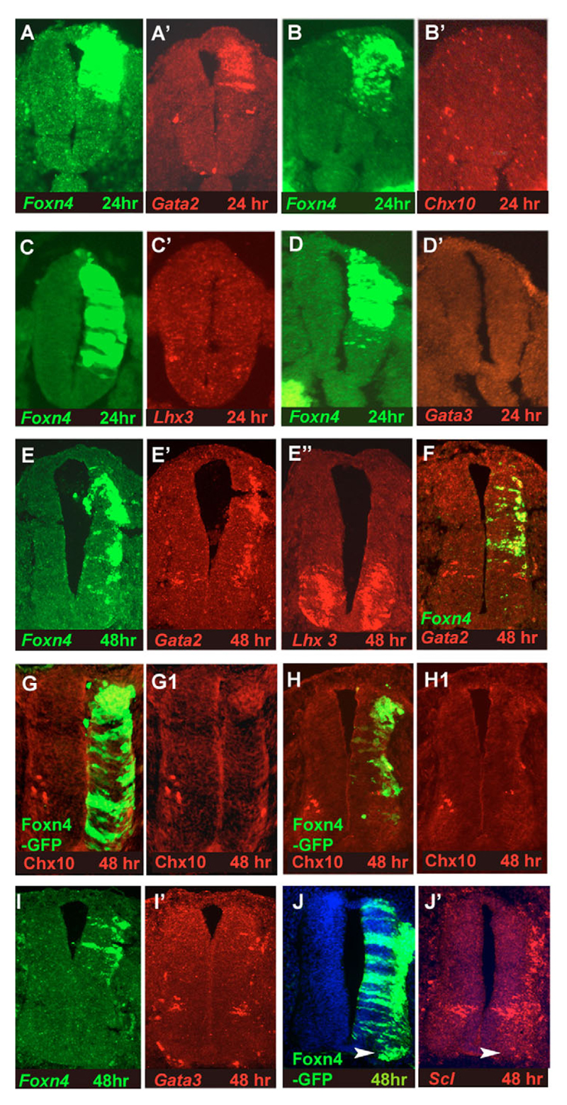
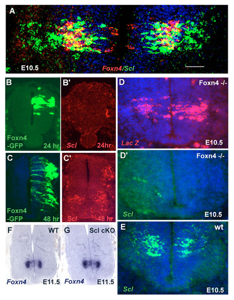
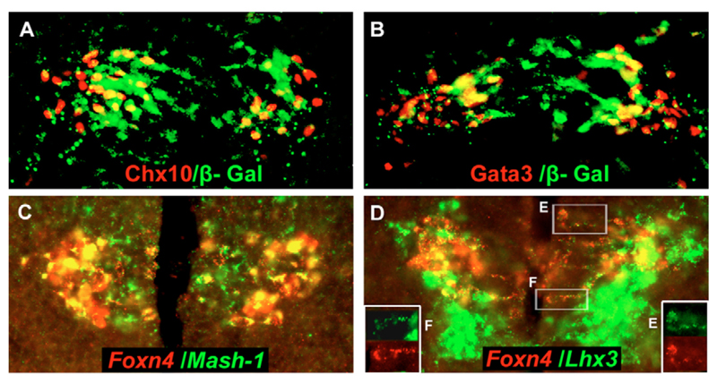
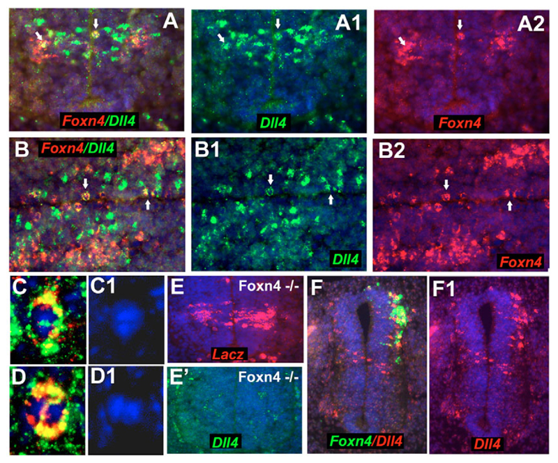
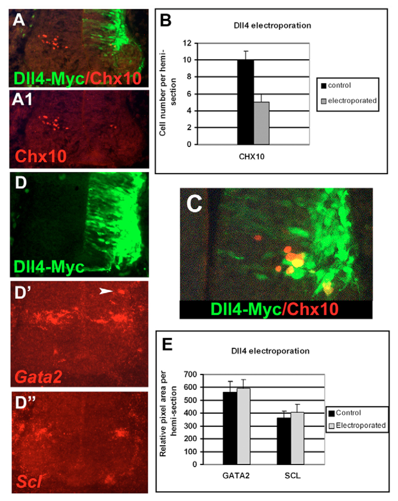
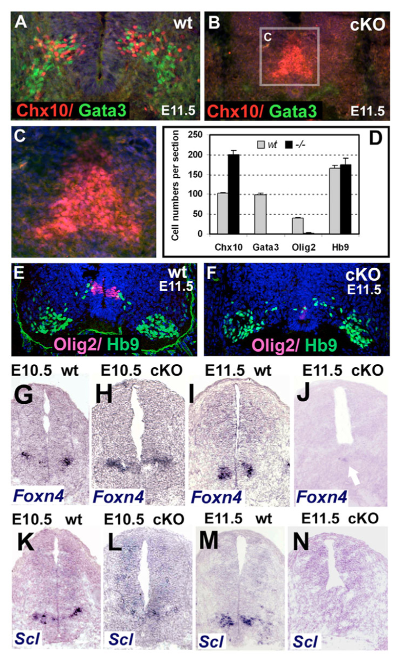
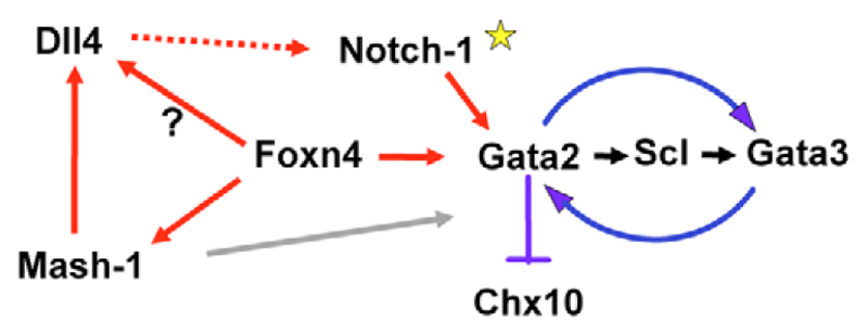
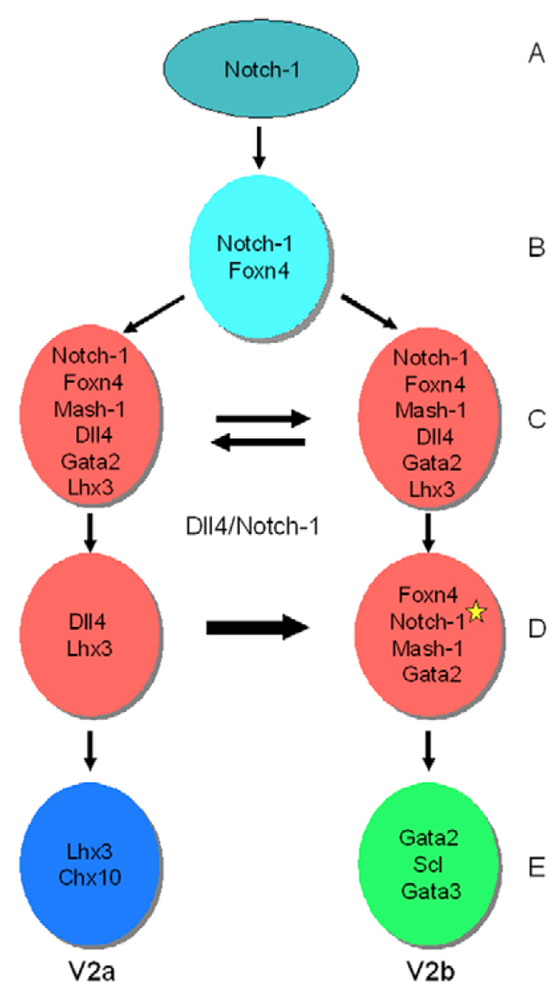
In the developing central nervous system, cellular diversity depends in part on organising signals that establish regionally restricted progenitor domains, each of which produces distinct types of differentiated neurons. However, the mechanisms of neuronal subtype specification within each progenitor domain remain poorly understood. The p2 progenitor domain in the ventral spinal cord gives rise to two interneuron (IN) subtypes, V2a and V2b, which integrate into local neuronal networks that control motor activity and locomotion. Foxn4, a forkhead transcription factor, is expressed in the common progenitors of V2a and V2b INs and is required directly for V2b but not for V2a development. We show here in experiments conducted using mouse and chick that Foxn4 induces expression of delta-like 4 (Dll4) and Mash1 (Ascl1). Dll4 then signals through Notch1 to subdivide the p2 progenitor pool. Foxn4, Mash1 and activated Notch1 trigger the genetic cascade leading to V2b INs, whereas the complementary set of progenitors, without active Notch1, generates V2a INs. Thus, Foxn4 plays a dual role in V2 IN development: (1) by initiating Notch-Delta signalling, it introduces the asymmetry required for development of V2a and V2b INs from their common progenitors; (2) it simultaneously activates the V2b genetic programme.


Figures

Fig. 1
Fig. 1. Foxn4 is sufficient to induce V2b and suppress V2a interneurons.
In this and subsequent figure legends, consecutive sections are labelled A,A',A" etc, and different fluorescence channels of the same micrograph are labelled A,A1,A2 etc. (A-J') Chick embryos were electroporated at st12-14 with β-actin-Foxn4-IRES-GFP and harvested after 24 or 48 hours. Expression of the vector was confirmed by in situ hybridisation (ISH) for Foxn4 or immunolabelling for GFP (panels marked Foxn4-GFP). Foxn4 induces robust ectopic expression of Gata2 at either 24 or 48 hours post-electroporation (A',E',F). Foxn4 induced Gata3 (D', I') and Scl (J', note ventral induction, arrowhead) only after 48 hours. Foxn4 does not induce ectopic expression of Chx10 (B',G,H) or Lhx3 (C',E"). On the contrary, Foxn4 represses endogenous Chx10 in the p2 domain (G,H).
Fig. 2
Fig. 2. Foxn4 lies upstream of Scl in V2b interneuron development.
(A) Double ISH for Scl (green) and Foxn4 (red). Confocal image of wild-type E10.5 mouse spinal cord, showing co-localisation of Scl and Foxn4 in some cells. (B-C') Sections from chicken embryos electroporated at st12-14 with β-actin-Foxn4-IRES-GFP and analysed after a further 24 (B) or 48 (C) hours. Foxn4 does not induce Scl after 24 hours (B'). At this stage, endogenous Scl is not expressed in the chick neural tube (B'). Foxn4 does induce ectopic Scl after 48 hours (C'). (D,D') Scl expression in the p2 domain is dependant on functional Foxn4. Consecutive sections from Foxn4-null mouse embryos at E10.5 were subjected to ISH for lacZ (D) or Scl (D'). The row of Scl-positive cells visible on the left of this section are endothelial cells. (E) Scl-positive cells in a wild-type mouse embryo at E10.5. (F,G) Foxn4 expression is not dependent on Scl. Foxn4 expression was visualised by ISH at E11.5 in wild-type (F) and Scl conditional null (G) mouse spinal cords (see Materials and methods). Scale bar: 20 μm.
Fig. 3
Fig. 3. Foxn4 is expressed in common precursors of V2a and V2b interneurons.
(A,B) Foxn4+/– mouse embryos were labelled by double immunohistochemistry for β-galactosidase (β-gal, green) and either Chx10 or Gata3 (red). Confocal microscopy reveals cells that are double labelled for β-gal and either Chx10 (A) or Gata3 (B), suggesting that Foxn4-expressing progenitors give rise to both V2a and V2b interneurons (INs). (C-F) Consistent with this conclusion, Foxn4-positive progenitors co-express Mash1 (C) and Lhx3 (D), markers that later segregate into V2b and V2a INs respectively. Individual Foxn4/Lhx3 double-positive cells (boxes E and F) are reproduced, with fluorescence channels separated, in the lower left and lower right corners, respectively, of D.
Fig. 4
Fig. 4. Foxn4 is necessary and sufficient to induce Dll4 in the p2 domain.
(A-D1) Double ISH for Dll4 (green) and Foxn4 (red) in wild-type E10.5 mouse embryos, counter-stained with Hoechst to visualise cell nuclei. A and B are transverse and longitudinal sections, respectively, of spinal cord. Foxn4 is expressed in some of the Dll4-positive cells within and outside the VZ (arrows). A significant proportion of double-labelled cells at the ventricular surface are pairs of cells in contact with each other, presumptive daughters of a recent progenitor cell division (e.g. arrows in B). Examples of these are shown at higher magnification in C,D; note the paired nuclei in C1,D1. (E,E') Foxn4-null mouse embryos at E10.5. (E) lacZ expression under Foxn4 control. (E') Dll4 expression in the p2 domain is abolished. (F,F1) Double ISH for Foxn4 (green) and Dll4 (red) showing that Foxn4 induces ectopic expression of Dll4 in electroporated st11-13 chick neural tube.
Fig. 5
Fig. 5. Dll4 inhibits V2a lineage progression.
Chick embryos were electroporated with human DLL4 (hDll4) in the form of hDll4-Myc at st14-16 and analysed after 48 hours. (A-B) Double immunolabelling for Chx10 (red) and hDll4-Myc (green) showing repression of Chx10-positive cells. (B) Quantification of Chx10-labelled cells showed a ~50% decrease on the hDll4-electroporated side compared with the contralateral, control side. (C) Some hDll4-electroporated cells co-express Chx10, consistent with the idea that Dll4 can suppress V2a generation in a non-cell-autonomous fashion. (D) Immunolabelling for hDll4-Myc (green). (D') Dll4 exceptionally can induce Gata2 (arrowhead). (D") Dll4 does not affect Scl expression. (E) Dll4 does not greatly affect generation of V2b INs, judging by ISH. Quantification of V2b markers Gata2 and Scl by pixel-counting software showed no significant effect on V2b production (see text for statistics).
Fig. 6
Fig. 6. Foxn4 controls Mash1 expression.
(A) Double ISH for Foxn4 (red) and Mash1 (green) in E10.5 mouse cord. Note the extensive overlap in the p2 domain. (B,B') Foxn4 induces ectopic expression of Cash1 in chick electroporation experiments. (C,D) Foxn4 expression does not depend on Mash1; there is no noticeable change in the Foxn4 ISH signal in Mash1-null mice compared with wild type. (E,E') Electroporation of β-actin-Mash1-IRES-GFP in the st13-14 chick neural tube induces Dll4 after 24 hours. Mash1 expression was confirmed by GFP immunolabelling (E) and Dll4 by ISH (E'). (F-G) Mash1 did not induce ectopic Chx10, but repressed endogenous Chx10 V2a INs in the p2 domain. G is a magnified view of the ventral part of panel F'. (H-H") Also, Mash1 did not induce ectopic V2b markers Gata2 or Scl. (I,J) Despite the fact that Mash1 is sufficient to induce Dll4 in chick (see E,E'), Mash1 is not required for Dll4 expression in mice; Dll4 is expressed as normal in the ventral spinal cord of Mash1-null mice.
Fig. 7
Fig. 7. Notch1 is required for specification of V2b interneurons.
Mice carrying a floxed allele of Notch1 and a Nestin-Cre transgene (Notch1 cKO mice) were analysed at E10.5 and E11.5 by ISH and double immunolabelling for V2a and V2b IN markers. (A-D) There is a two-fold increase in the number of Chx10 immunopositive V2a INs in the Notch1 cKO compared with wild type, whereas Gata3 immunopositive V2b INs are abolished. In addition, the Chx10-positive V2a INs accumulate near the midline of the spinal cord instead of migrating into the parenchyma. (E,F) Double immunolabelling for Olig2 (magenta) and Hb9 (green). In the Notch1 cKO, Olig2-positive cells are missing and the Hb9 population is similar to that in the control. Therefore, Notch1 activity is needed for V2b IN production; in the absence of Notch1, V2b INs are respecified as V2a INs with little or no influence on MN fate. (G-J) In the Notch1 cKO, expression of Foxn4 is increased at E10.5 relative to wild type (compare G with H), but is almost extinguished by E11.5 (I,J). (K-N) Scl (V2b INs) is reduced at E10.5 (K,L) and absent at E11.5 (M,N) in the Notch1 cKO. Note that the ventral half of the central canal (and the VZ) is lost in the Notch1 cKO mouse between E10.5 and E11.5.
Fig. 8
Fig. 8. Genetic interactions in the V2 interneuron lineage.
Red arrows represent positive intracellular interactions that we demonstrated in the present study, except for Foxn4 → (?) Dll4, which is speculative. Black arrows denote speculative interactions proposed in the present study. Blue arrows depict interactions demonstrated in previous studies (see below). The dashed red line represents the proposed intercellular Dll4/Notch1 interaction between sibling V2 progenitors, which results in Notch1 being activated (yellow star) in the cells that develop subsequently as V2b INs. The grey arrow signifies the requirement of Mash1 for proper V2b development (Li et al., 2005); this role of Mash1 is ill-defined (see Discussion). Mash1 is sufficient (in chick) but not necessary (in mice) for Dll4 upregulation (see Discussion). This diagram incorporates observations from a number of studies including the present one: Karunaratne et al. (Karunaratne et al., 2002) demonstrated reciprocal activation of Gata2 and Gata3 and repression of Chx10 by Gata2; Muroyama et al. (Muroyama et al., 2005) showed that Scl induces Gata2 and Gata3 and represses Chx10 in chick, and that Gata3 is abolished and Gata2 severely reduced in Scl-null mice; Li et al. (Li et al., 2005) showed that Mash1 expression is abolished in Foxn4-null mice.
Fig. 9
Fig. 9. Generation of V2a and V2b INs from common progenitors in the p2 domain.
Multipotent neuroepithelial (radial) progenitors (A), which do not express Foxn4, generate a population of V2a/V2b (p2) progenitors (B). All V2a/V2b progenitors express Foxn4, which induces the expression of Dll4, Gata2 and Mash1. These common progenitors also start to express Lhx3 at their final division (C). Notch1 is expressed in all p2 progenitors (Lindsell et al., 1996), so Notch1/Dll4 reciprocal cell-cell interactions are initiated (opposing arrows in C). This situation resolves into two populations of progenitors, one with activated Notch1 (Notch1*) and the other with Dll4 (D). Notch1* blocks the V2a fate and, in cooperation with Foxn4 and Mash1, specifies V2b IN fate (E). The complementary set of p2 progenitors (Dll4-positive) that fails to activate Notch1 adopts the V2a fate instead, possibly under the control of Lhx3 (Tanabe et al., 1998) (E). In this way, V2a and V2b INs are generated in a salt-and-pepper fashion during the same time window from a homogeneous population of p2 progenitors.

Similar articles

Cited by

References

    1. Artavanis-Tsakonas S, Rand MD, Lake RJ. Notch signaling: cell fate control and signal integration in development. Science. 1999;284:770–776. - PubMed
    1. Benedito R, Duarte A. Expression of Dll4 during mouse embryogenesis suggests multiple developmental roles. Gene Expr Patterns. 2005;5:750–755. - PubMed
    1. Briscoe J, Pierani A, Jessell TM, Ericson J. A homeodomain protein code specifies progenitor cell identity and neuronal fate in the ventral neural tube. Cell. 2000;101:435–445. - PubMed
    1. Casarosa S, Fode C, Guillemot F. Mash1 regulates neurogenesis in the ventral telencephalon. Development. 1999;126:525–534. - PubMed
    1. Castro DS, Skowronsky-Krawzyck D, Armant O, Donaldson IJ, Parras C, Hunt C, Critchley JA, Nguyen L, Gossler A, Göttgens B, et al. Proneural bHLH and Brn proteins coregulate a neurogenic program through cooperative binding to a conserved DNA motif. Dev Cell. 2006;11:831–844. - PubMed

Publication types

MeSH terms