. 2007 Aug 29;27(35):9262-9.
doi: 10.1523/JNEUROSCI.1843-07.2007.

Amyloid beta protein modulates glutamate-mediated neurotransmission in the rat basal forebrain: involvement of presynaptic neuronal nicotinic acetylcholine and metabotropic glutamate receptors

Affiliations

Amyloid beta protein modulates glutamate-mediated neurotransmission in the rat basal forebrain: involvement of presynaptic neuronal nicotinic acetylcholine and metabotropic glutamate receptors

James H Chin et al. J Neurosci. .

Abstract

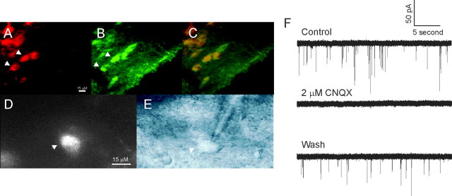
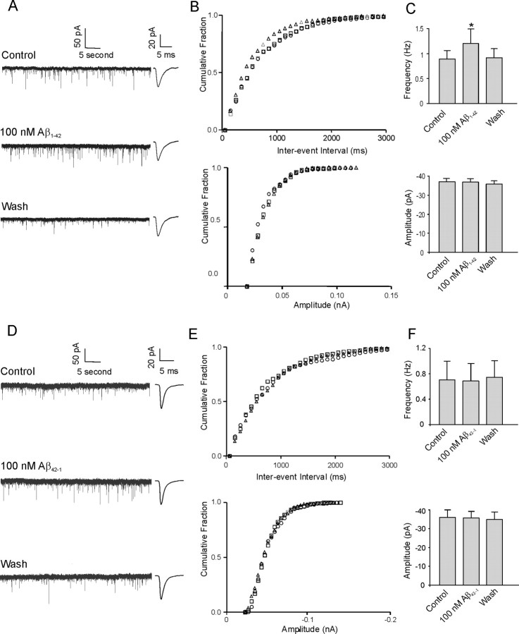
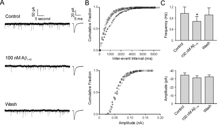
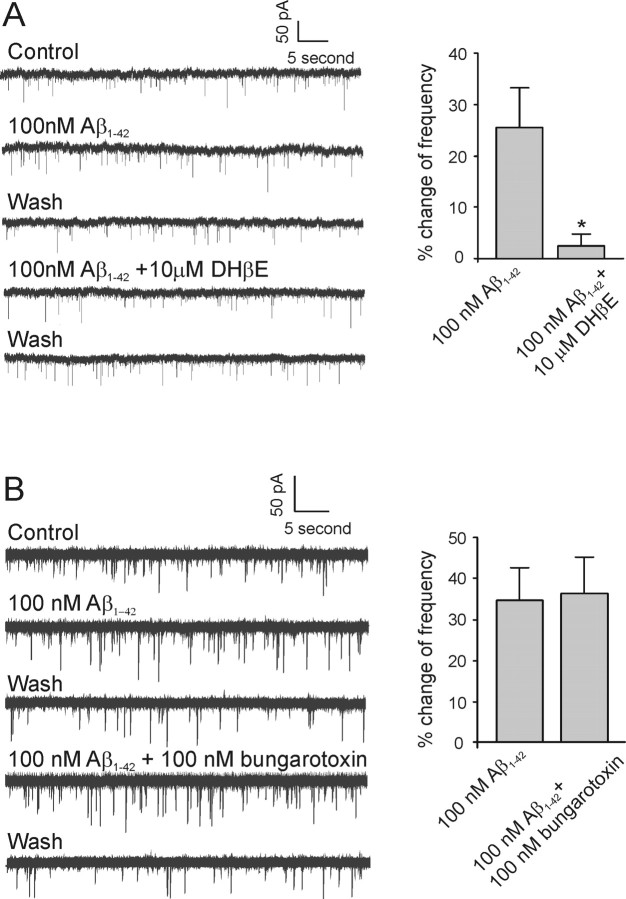
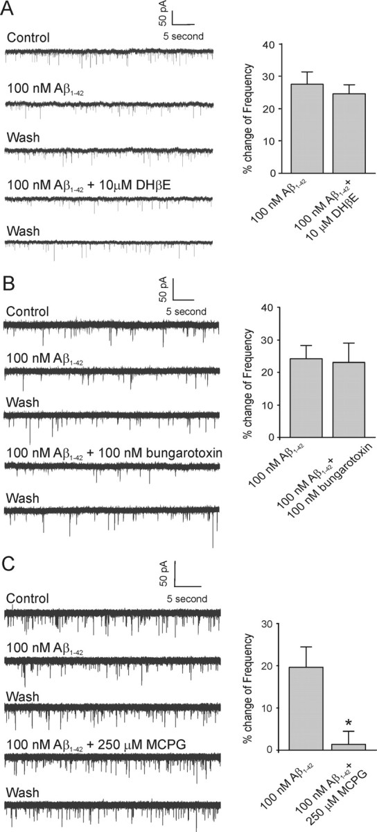
Amyloid beta (Abeta) protein, a 39-43 amino acid peptide deposited in brains of individuals with Alzheimer's disease (AD), has been shown to interact directly with a number of receptor targets including neuronal nicotinic acetylcholine receptors (nAChRs) and glutamate receptors. In this study, we investigated the synaptic effects of Abeta(1-42) on glutamate-mediated neurotransmission in the diagonal band of Broca (DBB), a cholinergic basal forebrain nucleus. Glutamatergic miniature EPSCs (mEPSCs) were recorded using whole-cell patch-clamp recordings from identified cholinergic DBB neurons in rat forebrain slices. In 54% of DBB neurons, bath application of Abeta(1-42) (100 nM), but not Abeta(42-1) (inverse fragment), significantly increased the frequency of mEPSCs without affecting amplitude or kinetic parameters (rise or decay time). In 32% of DBB neurons, bath application of Abeta(1-42) significantly decreased only the frequency but not amplitude of mEPSCs. Application of dihydro-beta-erythroidine (DHbetaE) (an antagonist for the alpha4beta2 subtype of nAChRs) but not alpha-bungarotoxin (an antagonist for the alpha7 subtype of nAChRs) blocked Abeta(1-42)-mediated increases in mEPSC frequency. The Abeta(1-42)-mediated increase in glutamatergic transmission is thus presynaptic and mediated via non-alpha7 AChRs. In contrast, Abeta(1-42)-mediated decreases in mEPSC frequency could not be antagonized by either DHbetaE or alpha-bungarotoxin. However, the Abeta(1-42)-evoked depression in mEPSC frequency was antagonized by (RS)-alpha-methyl-4-carboxyphenyglycine, a nonselective group I/II metabotropic glutamate receptor antagonist. These observations provide further insight into the mechanisms whereby Abeta affects synaptic function in the brain and may be relevant in the context of synaptic failure observed in AD.


Figures

Figure 1.
Figure 1.
Identification of cholinergic neurons and mEPSCs in the DBB. A, B, Horizontal slice of the rat forebrain containing the DBB viewed under a filter for Cy3-192 IgG (red) and a filter for Alexa 488 (green; vChAT). C, The overlay of images A and B shows that Cy3-192 IgG labels cells that are also vChAT positive (arrowheads in A, B). The calibration bar in A is the same for B and C. D, A Cy3-192 IgG-labeled cell in a slice viewed under a 60× water-immersion lens before recording (arrowhead). E, Differential infrared contrast image of the same cell viewed in C being recorded (arrowhead). The calibration bar in D is the same for E. F, mEPSC activity in neurons of DBB is reversibly abolished by CNQX, an AMPA/kainate receptor antagonist.
Figure 2.
Figure 2.
1–42, but not the reverse peptide Aβ42–1, increases mEPSC frequency but not amplitude in DBB neurons. A, Sample traces of mEPSCs under control conditions, during application of Aβ1–42, and during washout from a cholinergic DBB neuron. Insets beside each trace show averaged mEPSCs from 100 consecutive events under the three recording conditions. B, Cumulative probability analysis of mEPSCs of the same neuron as in A showing the distribution of the interevent interval (top) and peak amplitude (bottom) during control conditions (square), during application of 100 nm1–42 (triangle), and during washout (circle). Aβ1–42 caused a significant decrease in the interevent interval of mEPSCs without changing the distribution of the amplitude (p < 0.0005). C, Average frequency of mEPSCs (top) and average amplitude of mEPSCs (bottom) under control conditions, during application of Aβ1–42, and during washout (n = 15; *p < 0.05, significant difference from control). D, Sample traces of mEPSCs under control conditions, during application of Aβ42–1, and during washout from another cholinergic DBB neuron. Insets beside each trace show averaged mEPSCs from 100 consecutive events under the three recording conditions. E, The cumulative frequency histogram of an mEPSC interevent interval (top) and amplitude (bottom) from the same cell as in D demonstrates no significant change between control conditions (square) and during application of 100 nm42–1 (triangle). Washout is denoted by a circle. F, Average frequency of mEPSCs (top) and average amplitude of mEPSCs (bottom) during application of Aβ42–1 showing no significant difference between the peptide application and control or washout (n = 4; p > 0.05). Error bars indicate SEM.
Figure 3.
Figure 3.
1–42 also decreases mEPSC frequency but not amplitude in some DBB neurons. A, Sample traces of mEPSCs under control conditions, during application of Aβ1–42 and during washout from a cholinergic DBB neuron. Insets beside each trace show averaged mEPSCs from 100 consecutive events under the three recording conditions. B, Cumulative probability analysis of mEPSCs of the same neuron as in A showing the distribution of the interevent interval (top) and peak amplitude (bottom) during control conditions (square), during application of 100 nm1–42 (triangle), and during washout (circle). Aβ1–42 caused a significant increase in the interevent interval of mEPSCs without changing the distribution of the amplitude (p < 0.0005). C, Average frequency of mEPSCs (top) and average amplitude of mEPSCs (bottom) under control conditions, during application of Aβ1–42, and during washout (n = 9; *p < 0.05, significant difference from control). Error bars indicate SEM.
Figure 4.
Figure 4.
1–42-mediated increase in mEPSC frequency is antagonized by non-α7 but not α7 nAChR antagonists. A, Left, Sample traces of mEPSCs from a cholinergic DBB neuron in response to Aβ1–42 and a combination of Aβ1–42 and DHβE (a non-α7 nAChR antagonist), respectively. Right, Histograms depicting the average effect of 10 μm DHβE on the Aβ1–42-mediated increase in mEPSC frequency (n = 5; *p < 0.05, significant difference from control). B, Left, Sample traces of mEPSCs from another cholinergic DBB neuron in response to Aβ1–42 and a combination of Aβ1–42 and α-bungarotoxin (α7 nAChR antagonist), respectively. Right, Histograms depicting the average effect of 100 nm α-bungarotoxin on the Aβ1–42-mediated increase in mEPSC frequency (n = 8; p > 0.05). Error bars indicate SEM.
Figure 5.
Figure 5.
1–42-mediated decrease in mEPSC frequency is not antagonized by nAChR antagonists but by mGluR antagonists. A, Left, Sample traces of mEPSCs from a cholinergic DBB neuron in response to Aβ1–42 and a combination of Aβ1–42 and DHβE (non-α7 nAChR antagonist), respectively. Right, Histograms depicting the average effect of DHβE on the Aβ1–42-mediated decrease in mEPSC frequency (n = 5; p > 0.05). B, Left, Sample traces of mEPSCs from another cholinergic DBB neuron in response to Aβ1–42 and a combination of Aβ1–42 and α-bungarotoxin (α7 nAChR antagonist), respectively. Right, Histograms depicting the average effect of α-bungarotoxin on the Aβ1–42-mediated decrease in mEPSC frequency (n = 4; p > 0.05). C, Left, Sample traces of mEPSCs from another cholinergic DBB neuron in response to Aβ1–42 and a combination of Aβ1–42 and MCPG (nonselective group I/group II mGluR antagonist), respectively. Right, Histograms depicting the average effect of DHβE on the Aβ1–42-mediated decrease in mEPSC frequency (n = 4; *p < 0.05, significant difference from control). Error bars indicate SEM.

References

    1. Alkondon M, Rocha ES, Maelicke A, Albuquerque EX. Diversity of nicotinic acetylcholine receptors in rat brain. α-Bungarotoxin-sensitive nicotinic receptors in olfactory bulb neurons and presynaptic modulation of glutamate release. J Pharmacol Exp Ther. 1996;278:1460–1471. - PubMed
    1. Anwyl R. Metabotropic glutamate receptors: electrophysiological properties and role in plasticity. Brain Res Brain Res Rev. 1999;29:83–120. - PubMed
    1. Ashenafi S, Fuente A, Criado JM, Riolobos AS, Heredia M, Yajeya J. Beta-amyloid peptides25–35 depresses excitatory synaptic transmission in the rat basolateral amygdala “in vitro”. Neurobiol Aging. 2005;26:419–428. - PubMed
    1. Blanchard BJ, Thomas VL, Ingram VM. Related mechanism of membrane depolarization caused by the Alzheimer Abeta1–42 peptide. Biochem Biophys Res Commun. 2002;293:1197–1203. - PubMed
    1. Clements JD, Bekkers JM. Detection of spontaneous synaptic events with an optimally scaled template. Biophys J. 1997;73:220–229. - PMC - PubMed

Publication types

MeSH terms