. 2007 Aug 29;27(35):9380-91.
doi: 10.1523/JNEUROSCI.5092-06.2007.

Differential abilities of SNAP-25 homologs to support neuronal function

Affiliations

Differential abilities of SNAP-25 homologs to support neuronal function

Ignacio Delgado-Martínez et al. J Neurosci. .

Abstract

The SNAP receptor (SNARE) complex, consisting of synaptosome-associated protein of 25 kDa (SNAP-25), synaptobrevin-2, and syntaxin-1, is involved in synaptic vesicles exocytosis. In addition, SNAP-25 has been implicated in constitutive exocytosis processes required for neurite outgrowth. However, at least three isoforms of SNAP-25 have been reported from neurons: SNAP-23, which is also present in non-neuronal cells, and the two alternative splice variants SNAP-25a and SNAP-25b. Here, we studied the differential ability of these isoforms to support the functions previously broadly ascribed to "SNAP-25." We studied the rescue of snap-25 null neurons in culture with different SNAP-25 homologs. We find that deletion of SNAP-25 leads to strongly reduced neuron survival, and, in the few surviving cells, impaired arborization, reduced spontaneous release, and complete arrest of evoked release. Lentiviral expression of SNAP-25a, SNAP-25b, or SNAP-23 rescued neuronal survival, arborization, amplitude, and frequency of spontaneous events. Also evoked release was rescued by all isoforms, but synchronous release required SNAP-25a/b in both glutamatergic and GABAergic neurons. SNAP-23 supported asynchronous release only, reminiscent of synaptotagmin-1 null neurons. SNAP-25b was superior to SNAP-25a in vesicle priming, resembling the shift to larger releasable vesicle pools that accompanies synaptic maturation. These data demonstrate a differential ability of SNAP-25b, SNAP-25a, and SNAP-23 to support neuronal function.


Figures
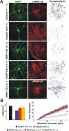
Figure 1.
Figure 1.
Elimination of SNAP-25 leads to impaired neuronal survival and outgrowth. A, Double staining for SNAP-25 (or SNAP-23) and synaptophysin as a synaptic marker of primary cultured hippocampal neurons infected with recombinant lentiviruses. A synapsin promotor was used to restrict eGFP expression to neurons and enable morphological analysis (left column). This revealed inferior outgrowth/branching in Snap-25 null (−/−) neurons compared with control (+/+; +/−). Nevertheless, neurons lacking SNAP-25 still formed synaptophysin-positive synapses. Expression of SNAP-25a, SNAP-25b, or SNAP-23 in Snap-25 null neurons recovered the morphology. B, Left, The number of cells (mean ± SEM) present after 10–14 d in culture. Survival of null neurons was dramatically reduced but rescued with SNAP-25a, SNAP-25b, or SNAP-23 expression. Right, The number of branches (mean ± SEM) as a function of distance to the soma. Neurite extension in surviving Snap-25 null neurons was significantly depressed (***p = 0.001, Student's t test) compared with control neurons or null neurons rescued with SNAP-25a, SNAP-25b, or SNAP-23.
Figure 2.
Figure 2.
Localization of SNAP-25 isoforms. A, Neurons (from wild-type animals) expressing GFP–SNAP-25a/b or GFP–SNAP-23 were fixed and stained with anti-GFP and Alexa-488 to increase the fluorescence in the green channel. The neurons were costained with anti-synaptophluorin, which was imaged in the red channel (right column). All SNAP-25 isoforms were present throughout the neuritic tree and overlapped with the synaptic marker. B, GFP fluorescence (± SEM) in live (unfixed) neurons expressing GFP–SNAP-23 (SN23; n = 55 cells), GFP–SNAP-25a (SN25a; n = 50 cells), and GFP–SNAP-25b (SN25b; n = 55 cells). The fluorescence was measured in the cell body and indicates similar expression levels.
Figure 3.
Figure 3.
Stimulus-dependent recycling of synaptic vesicles requires a SNAP-25 isoform. A–E, Examples of control hippocampal neurons stained with 400 APs (A), Snap-25 null neurons (B), and null neurons rescued with SNAP-25a (C), SNAP-25b (D), and SNAP-23 (E). Staining was not possible in the absence of SNAP-25, indicating a lack of synaptic vesicle recycling. Scale bars, 10 μm. F, The intensity of FM 5-95 staining by 40 AP and 400 AP loading. The background intensity after full destaining was subtracted. G, Destaining (mean ± SEM) of synaptic boutons under strong electrical stimulation (3 pulses of 400 AP at 10 Hz) after loading with 400 AP. Color coding as above. The destaining kinetics was indistinguishable between groups.
Figure 4.
Figure 4.
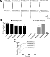
SNAP-25 supports synchronous and SNAP-23 asynchronous release. A, Autaptic EPSCs in neurons expressing SNAP-25a, SNAP-25b, or SNAP-23 compared with eGFP-expressing and uninfected neurons. Control (+/+; +/−) neurons are shown in black, and snap-25 null neurons are shown in gray. No evoked responses were found in the absence of SNAP-25. Strikingly, snap-25 null neurons expressing SNAP-23 produced evoked responses lacking the fast synchronous component. B, Mean ± SEM. EPSC amplitudes for the groups described above. SNAP-25a rescue led to smaller EPSC amplitudes than SNAP-25b rescue or control. **p < 0.01; ***p < 0.001. C, Integrated EPSCs for control and rescued neurons. SNAP-23 rescued neurons presented slower release of vesicles. For kinetic parameters, see Table 1. WT, Wild type.
Figure 5.
Figure 5.
All SNAP-25 isoforms rescue the sucrose pool. A, Example recordings of 500 mm sucrose application in control hippocampal neurons (+/+; +/−), Snap-25 null neurons (−/−), and after rescue with SNAP-25a, SNAP-25b, and SNAP-23. A small vesicle pool was released by sucrose even in knock-out neurons. B, Mean ± SEM values of the “sucrose pool” for each of the groups described above. ***p < 0.001.
Figure 6.
Figure 6.
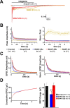
High-frequency stimulation reveals differences between SNAP-25a, SNAP-25b, and SNAP-23. A, Example traces of high-frequency train stimulation (100 AP at 40 Hz) of snap-25 null neurons (gray), SNAP-25b rescued neurons (red), and neurons rescued with SNAP-23 (yellow). Stimulation artifacts have been removed. B, Mean ± SEM peak current amplitudes (left) and after normalization to the first stimulation (right) during high-frequency train stimulation. SNAP-23 expression in null neurons caused build-up of currents attributable to overlap of asynchronous release components during the train. C, Mean ± SEM synchronized EPSC amplitude (left) and after normalization to the first stimulation (right) during high-frequency train stimulation. Depression during the train was indistinguishable between SNAP-25a and SNAP-25b. D, Left, Example cumulative trace of the EPSC amplitudes during the 40 Hz train stimulation. The data between 1 and 2.5 s were fitted with a straight line (blue). The line was back-extrapolated to 0 to calculate the RRP size as determined by RRPer. Right, Mean ± SEM of the RRPer values calculated as shown to the left. RRPer was significantly smaller when SNAP-25a was used for rescuing null neurons instead of SNAP-25b. **p < 0.01.
Figure 7.
Figure 7.
Smaller and fewer spontaneous events in the Snap-25 null neurons are rescued by SNAP-25 and SNAP-23. A, Example averaged mEPSCs from single cells. Events were indistinguishable in shape; however, they were smaller in snap-25 null neurons. B, Mean ± SEM values of mEPSC amplitudes. The mEPSC size was significantly reduced in Snap-25 null neuron. ***p < 0.001.
Figure 8.
Figure 8.
SNAP-25 is essential for fast release in GABAergic neurons. A, Example recordings of GABAergic responses in striatal neurons from control (+/+; +/−), snap-25 null neurons, and null neurons rescued with SNAP-25a, SNAP-25b, and SNAP-23. No IPSCs were found in null, suggesting that inhibitory neurons need SNAP-25 for synaptic transmission. Null neurons rescued by SNAP-25a or SNAP-25b evoked normal IPSCs. Responses in SNAP-23 rescued neurons were smaller and the fast component was abolished, mimicking the situation in glutamatergic neurons. B, Example of a Snap-25 null neuron rescued with SNAP-25b showing (left) reversal of the IPSC after changing the holding potential from −100 mV (black) to −20 mV (light gray) in 10 mV steps and blockage by the GABAA antagonist bicuculline (right). C, Left, Mean ± SEM values of the IPSC amplitudes. Right, Example of the integrated charge released by the IPSC for each of the groups. The kinetics of evoked release for SNAP-23 rescued neurons was slower than for the other groups. ***p < 0.001.

References

    1. Arantes RM, Andrews NW. A role for synaptotagmin VII-regulated exocytosis of lysosomes in neurite outgrowth from primary sympathetic neurons. J Neurosci. 2006;26:4630–4637. - PMC - PubMed
    1. Bark C, Bellinger FP, Kaushal A, Mathews JR, Partridge LD, Wilson MC. Developmentally regulated switch in alternatively spliced SNAP-25 isoforms alters facilitation of synaptic transmission. J Neurosci. 2004;24:8796–8805. - PMC - PubMed
    1. Bark IC. Structure of the chicken gene for SNAP-25 reveals duplicated exon encoding distinct isoforms of the protein. J Mol Biol. 1993;233:67–76. - PubMed
    1. Bark IC, Hahn KM, Ryabinin AE, Wilson MC. Differential expression of SNAP-25 protein isoforms during divergent vesicle fusion events of neural development. Proc Natl Acad Sci USA. 1995;92:1510–1514. - PMC - PubMed
    1. Bekkers JM, Stevens CF. Excitatory and inhibitory autaptic currents in isolated hippocampal neurons maintained in cell culture. Proc Natl Acad Sci USA. 1991;88:7834–7838. - PMC - PubMed

Publication types

MeSH terms

Substances