. 2007 Aug 29;27(35):9513-24.
doi: 10.1523/JNEUROSCI.1493-07.2007.

Isoform-selective susceptibility of DISC1/phosphodiesterase-4 complexes to dissociation by elevated intracellular cAMP levels

Affiliations

Isoform-selective susceptibility of DISC1/phosphodiesterase-4 complexes to dissociation by elevated intracellular cAMP levels

Hannah Murdoch et al. J Neurosci. .

Abstract

Disrupted-in-schizophrenia 1 (DISC1) is a genetic susceptibility factor for schizophrenia and related severe psychiatric conditions. DISC1 is a multifunctional scaffold protein that is able to interact with several proteins, including the independently identified schizophrenia risk factor phosphodiesterase-4B (PDE4B). Here we report that the 100 kDa full-length DISC1 isoform (fl-DISC1) can bind members of each of the four gene, cAMP-specific PDE4 family. Elevation of intracellular cAMP levels, so as to activate protein kinase A, caused the release of PDE4D3 and PDE4C2 isoforms from fl-DISC1 while not affecting binding of PDE4B1 and PDE4A5 isoforms. Using a peptide array strategy, we show that PDE4D3 binds fl-DISC1 through two regions found in common with PDE4B isoforms, the interaction of which is supplemented because of the presence of additional PDE4B-specific binding sites. We propose that the additional binding sites found in PDE4B1 underpin its resistance to release during cAMP elevation. We identify, for the first time, a functional distinction between the 100 kDa long DISC1 isoform and the short 71 kDa isoform. Thus, changes in the expression pattern of DISC1 and PDE4 isoforms offers a means to reprogram their interaction and to determine whether the PDE4 sequestered by DISC1 is released after cAMP elevation. The PDE4B-specific binding sites encompass point mutations in mouse Disc1 that confer phenotypes related to schizophrenia and depression and that affect binding to PDE4B. Thus, genetic variation in DISC1 and PDE4 that influence either isoform expression or docking site functioning may directly affect psychopathology.


Figures

Figure 1.
Figure 1.
Interaction of the fl-DISC1 isoform with PDE4 isoforms from all four PDE4 gene families. An N-terminally FLAG-tagged form of the fl-DISC1 isoform (FlagDISC1) was coexpressed in HEK293 cells with PDE4 isoform PDE4A5 (A), PDE4B1 (B), PDE4C2 (C), or PDE4D3 (D). Cells were challenged with vehicle or 100 μm forskolin (Fsk)/100 μm IBMX for 15 min before harvesting. Equalized amounts of cell lysate were immunoprecipitated (IP) with either control IgG or M2 anti-FLAG antibody and immunoblotted (IB) with PDE4A (A), PDE4B (B), PDE4C (C), or PDE4D (D) antisera (top). Equal immunocapture of FlagDISC1 was determined by immunoblotting with the anti-FLAG M2 antibody (bottom). The relative expression levels of FlagDISC1 and PDE4 isoforms in ∼5% of total cell lysate input used for coimmunoprecipitation assays were determined by direct immunoblotting with the PDE4 antisera (top) and the anti-FLAG M2 antibody (bottom).
Figure 2.
Figure 2.
PDE4B isoforms remain stably associated with the fl-DISC1 isoform; DISC1 interaction with members of the PDE4D subfamily are disrupted by treatment with forskolin/IBMX. A, HEK293 cells were cotransfected to express Flag-tagged fl-DISC1 and PDE4B isoforms PDE4B1, PDE4B2, and PDE4B3. Cells were challenged by treatment with vehicle or 100 μm forskolin/100 μm IBMX for 15 min. Equal amounts of cell lysate were subjected to anti-FLAG M2 immunoprecipitation. The coprecipitation of PDE4B1, PDEB2, or PDEB3 with FlagDISC1 was detected using the PDE4B antibody (top). Equalized FlagDISC1 expression was confirmed by immunoblotting with the anti-FLAG M2 antibody (bottom). B, HEK293 cells coexpressing Flag-tagged fl-DISC1 and PDE4D isoforms PDE4D3 and PDE4D5 were challenged with vehicle or 100 μm forskolin/100 μm IBMX for 15 min. Cell lysates were immunoprecipitated with the PDE4D antibody. Coprecipitated FlagDISC1 was visualized using the anti-FLAG M2 antibody (top). The relative expression levels of FlagDISC1 and the PDE4D isoforms in cell lysates were determined by immunoblotting with the anti-FLAG M2 (middle) and PDE4D (bottom) antibodies. C, SHSY5Y neuroblastoma cells were cotransfected with Flag-tagged fl-DISC1 and either PDE4B1 or PDE4D3. Cells were treated with or without 100 μm forskolin/100 μm IBMX for 15 min before preparation of cell lysates. Anti-FLAG immunoprecipitation was performed to isolate DISC1 protein complexes. The amount of PDE4B1 and PDE4D3 present in FlagDISC1 immunoprecipitates (top) and expression in cell lysates (bottom) was determined by immunoblotting with the PDE4B and PDE4D antibodies, respectively. D, SHSY5Y cells were treated with or without 100 μm forskolin/100 μm IBMX for 15 min before preparation of cell lysates. Anti-PDE4B immunoprecipitation was performed to isolate endogenous PDE4B–DISC1 complexes. The amount of PDE4B (bottom) and DISC1 (top) present in PDE4B immunoprecipitates was determined by immunoblotting. An antibody that specifically recognizes the 100 kDa fl-DISC1 isoform was used (Ogawa et al., 2005). Fsk, Forskolin; IP, immunoprecipitated; IB, immunoblotted.
Figure 3.
Figure 3.
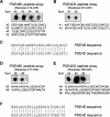
Probing DISC1 peptide arrays for PDE4B and PDE4D interaction sites. A, B, An array of immobilized peptide spots of overlapping 25-mer peptides, each shifted along by 5 aa in the entire sequence of the fl-DISC1 isoform, was probed for interaction with MBP fusion proteins of PDE4B1 (MBP-4B1) or PDE4D3 (MBP-4D3) or MBP alone and detected by immunoblotting. Dark spots were evidence of positive interaction, whereas noninteracting peptides left white (blank) spots. In all other sections of the array, spots were blank with the probes. Spot numbers relate to the 25-mer peptides in the array with the amino acid sequence of the peptide spots given below the array data. C, Schematic of DISC1 and PDE4 protein structure. DISC1 comprises an N-terminal globular domain with predicted coiled-coil (CC) regions within its C-terminal domain. Long PDE4 isoforms consist of two upstream conserved regions (UCR1 and UCR2) linked to the CAT subunit by linker regions. Distinct PDE4 isoforms are characterized by a unique N-terminal region. The sites of DISC1–PDE4 interaction established from peptide array analyses are highlighted (cs, common PDE4 binding site; 4bss, PDE4B-specific binding site). The relative position of mutations in mouse Disc1 that confer phenotypes related to psychiatric illness are indicated, namely Q31L and L100P D, DISC1 peptide arrays were probed with either MBP-4B1 or a GST fusion protein of PDE4B2 (GST-4B2), and interaction was detected by immunoblotting. Positively interacting peptide spots are shown with the related DISC1 regions given. E, HEK293 cells were transfected with the indicated pEBG-2T DISC1 plasmids and FLAG-tagged PDE4B3. Lysates were incubated with glutathione–Sepharose beads to capture GST or GST–fl-DISC1 complexes and immunoblotted with the anti-GST antibody to detect fl-DISC1 fusion proteins (data not shown) or anti-FLAG M2 antibody to detect the presence of coprecipitated PDE4B3. F, An MBP fusion protein of PDE4B1 was used to probe peptides reflecting the murine Disc1 sequence, namely one containing Gln31 (Q31), GHGLPPAVAPQ(31)RRRLTRRPGYMRST (left) and one containing Leu100 (L100), SHCQASLVGKPFL(100)KSSLVPAVASEG (right). In addition the indicated mutant peptides were probed to assess PDE4B1 binding to Leu100Ala, Leu100Pro, Gln31Ala, and Gln31Leu. G, Densitometric analysis of MBP–PDE4B1 interaction with the wild-type and indicated mutant forms of the Leu100 SHCOASLVGKPFL(100)KSSLVPAVASEG peptide. H, GST fusion proteins of individual PDE4D domains, UCR1, UCR2, and CAT were used to probe fl-DISC1 peptide arrays and binding of probes detected by immunoblotting with the anti-GST antibody. The interactions of the PDE4D probes with fl-DISC1 peptide spots of the common PDE4 binding sites (as determined in Fig. 4A) are shown. I, HEK293 cells coexpressing Flag-tagged fl-DISC1 and PDE4D3 were incubated for 2–3 h with 50 μm stearoylated DISC1 peptides of DISC1 regions encompassing cs1 (CS1P, SHSAFTSSFSFIRLSLGSAGERGE) and cs2 (CS2P, IRSLTSEREGLEGLLSKLLVLSSRN). Cells were then harvested, and lysates were immunoprecipitated with PDE4D antisera. Coprecipitation of FlagDISC1 was assessed by immunoblotting with the anti-FLAG M2 antibody (top). Equal immunocapture of PDE4D3 was determined by immunoblotting immunoprecipitates with the PDE4D antibody (middle). The relative expression level of FlagDISC1 in ∼5% of total cell lysate input used for coimmunoprecipitation assays was determined by direct immunoblotting with the anti-FLAG M2 antibody (bottom). J, As in I, cells coexpressing Flag-tagged fl-DISC1 and PDE4B1 were incubated with stearoylated DISC1 peptides CS1P and CS2P. Cell lysates were then incubated with PDE4B antisera, and PDE4B immunoprecipitates were immunoblotted with anti-FLAG M2 antibody to detect coprecipitated FlagDISC1 (top). Immunocapture of PDE4B1 was confirmed by immunoblotting immunoprecipitates with PDE4B antisera (middle). Equivalent input expression of FlagDISC1 was determined by immunoblotting ∼5% of total cell lysate used for the coimmunoprecipitation assays with the anti-FLAG M2 antibody (bottom). IP, Immunoprecipitated; IB, immunoblotted.
Figure 4.
Figure 4.
Probing PDE4B and PDE4D peptide arrays for fl-DISC1 interaction sites: PDE4 subfamilies exhibit common regions of DISC1 binding. A, B, D, E, Peptide arrays of the PDE4B (A, D) and PDE4D (B, E) sequences were probed with partially purified [35S]-labeled fl-DISC1 protein, and binding to peptide spots was detected by phosphorimager analysis. C, F, Sites common to both PDE4B and PDE4D for fl-DISC1 interaction are shown with the sequence homology between PDE4B and PDE4D at these binding regions highlighted.
Figure 5.
Figure 5.
Probing PDE4B and PDE4D peptide arrays for DISC1 interaction sites: PDE4B- and PDE4D-specific sites of DISC1 binding. A, B, D, E, As in Figure 4, PDE4B (A, E) and PDE4D (B, D) peptide arrays were probed with [35S]-fl-DISC1. C, F, Sites of [35S]-DISC1 binding unique to PDE4B or PDE4D are shown with comparative PDE4B and PDE4D sequence alignment at these sites highlighted.
Figure 6.
Figure 6.
N-terminal truncation of fl-DISC1 sensitizes PDE4B1 to cAMP-mediated release. HEK293 cells cotransfected with PDE4B1 and either myc-HIS-tagged fl-DISC1 or myc-HIS-tagged DISC1 N-terminal truncate lacking residues 1–187 were treated with vehicle or 100 μm forskolin/100 μm IBMX for 15 min before harvesting and immunoprecipitation with the PDE4B antibody. Coprecipitated myc-HIS-DISC1 species were detected with the anti-myc 9E10 antibody (top). Successful immunocapture of PDE4B1 was demonstrated with the PDE4B antibody (bottom). The input signal shows the expression levels of PDE4B1 (bottom) and myc-HIS DISC1 proteins (top) as determined by direct immunoblotting of ∼5% total cell lysate used for immunoprecipitation assays. IP, Immunoprecipitated; IB, immunoblotted.
Figure 7.
Figure 7.
Activation of PKA mediates PDE4D3 release from the fl-DISC1 isoform. A, HEK293 cells exogenously expressing Flag-tagged fl-DISC1, PDE4D3 and EPAC1 were challenged with either vehicle, 100 μm forskolin/100 μm IBMX or EPAC agonist (8-pCPT-2′-O-Me-cAMP, 10 μm) for 15 min. FlagDISC1 complexes were isolated from cell lysates by immunoprecipitation with the anti-FLAG M2 antibody, and the PDE4D antibody was used to detect coprecipitated PDE4D3 (top). Equalized capture of FlagDISC1 in immunoprecipitates was determined with the anti-FLAG M2 antibody (bottom). The input signals show relative levels of FlagDISC1 (top) and PDE4D3 (bottom) expression in ∼5% of the total cell lysate used for immunoprecipitation experiments. Cellular activation of EPAC1 was visualized by immunofluorescent staining of cells with antisera specific for the detection of cAMP-activated EPAC1. Confocal imaging of cells immunostained with the cAMP-activated EPAC1 antibody are shown. B, HEK293 cells were transfected to express either Flag-tagged fl-DISC1, PDE4D3, and empty vector or Flag-tagged fl-DISC1, PDE4D3, and PKI. Cells were then treated with or without 100 μm forskolin/100 μm IBMX for 15 min. After harvesting, equal amounts of cell lysate were incubated with the anti-FLAG M2 antibody to isolate FlagDISC1 complexes. FlagDISC1 immunoprecipitates were immunoblotted with the PDE4D (top) and anti-FLAG M2 (bottom) antibodies. Relative expression of PDE4D3 and FlagDISC1 in ∼ 5% of cell lysate input for immunoprecipitation experiments is shown in the top and bottom panels, respectively. The cells imaged in A, as determined by bright-field microscopy, are shown. C, HEK293 cells coexpressing Flag-tagged fl-DISC1 and PDE4D3 were treated with vehicle, 100 μm forskolin/100 μm IBMX, or 200 ng/ml EGF for 15 min. FlagDISC1 complexes were immunopurified with the anti-FLAG M2 antibody, and the PDE4D antibody was used to detect coprecipitated PDE4D3 (top). Equalized capture of FlagDISC1 in immunoprecipitates was determined with the anti-FLAG M2 antibody (bottom). The input signals show relative levels of FlagDISC1 (top) and PDE4D3 (bottom) expression in ∼5% of the total cell lysate used for immunoprecipitation experiments. EGF-mediated Erk activation was confirmed by immunoblotting of cell lysates with phospho-Erk1/2 antisera (top right). IP, Immunoprecipitated; IB, immunoblotted.
Figure 8.
Figure 8.
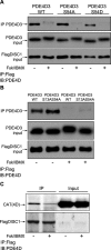
PKA phosphorylation of PDE4D3 is not responsible for PKA-induced dissociation of PDE4D3 from the fl-DISC1 isoform. A, HEK293 cells cotransfected with Flag-tagged fl-DISC1 and either wild-type PDE4D3 or PDE4D3 mutants S54A or S54D were challenged with or without 100 μm forskolin/100 μm IBMX. Equal amounts of cell lysate were used to prepare anti-FLAG M2 immunoprecipitates, and coprecipitated PDE4D3 was detected by immunoblotting with the PDE4D antibody (top). Equalized expression of PDE4D3 and FlagDISC1 in input lysates for immunoprecipitation assays was verified by immunoblotting with the PDE4D (middle) and anti-FLAG M2 (bottom) antibodies. B, Similar to above, HEK293 cells coexpressing Flag-tagged fl-DISC1 with either wild-type PDE4D3 or PDE4D3 S13A:S54A mutant were treated with or without forskolin/IBMX for 15 min. Cell lysates were then subjected to anti-FLAG M2 immunoprecipitation, and the coprecipitated PDE4D3 was detected with the PDE4D antibody (top). Relative expression inputs of PDE4D3 and FlagDISC1 are shown in the middle and bottom panels, respectively. C, HEK293 cells were cotransfected with Flag-tagged fl-DISC1 and PDE4D CAT domain and treated with vehicle or 100 μm forskolin/100 μm IBMX for 15 min. Anti-FLAG M2 antibody was used to immunoprecipitate FlagDISC1, and coprecipitating CAT was visualized by immunoblotting with the PDE4D antibody (top). Equalized expression of CAT and FlagDISC1 in input lysates for immunoprecipitation assays was verified by immunoblotting with the PDE4D (top) and anti-FLAG M2 (bottom) antibodies. IP, Immunoprecipitated; IB, immunoblotted; WT, wild type.
Figure 9.
Figure 9.
cAMP-induced PKA activation does not elicit PKA phosphorylation of DISC1 in vivo. Attenuation of DISC1:PDE4D3 interaction is not mediated through PKA phosphorylation of DISC1. A, COS1 cells coexpressing Flag-tagged fl-DISC1 plus empty vector or Flag-tagged fl-DISC1 plus PKI were metabolically labeled with [32P]orthophosphate and challenged with vehicle or 100 μm forskolin/100 μm IBMX before harvesting. Equivalent amounts of cell lysate were incubated with anti-FLAG M2 antibody to immunoprecipitate Flag-tagged DISC1. Immunoprecipitates were resolved by SDS-PAGE, and radiolabeled Flag-tagged DISC1 was visualized by autoradiography. B, HEK293 cells coexpressing PDE4D3 with either Flag-tagged fl-DISC1 or the Flag-tagged-fl-DISC1 T744A mutant were treated with either vehicle or 100 μm forskolin/100 μm IBMX for 15 min. Equalized cell lysates were incubated with the PDE4D antibody to precipitate PDE4D3, and coprecipitated FlagDISC1 was determined by immunoblotting with the anti-FLAG M2 antibody (top). Immunocapture of PDE4D3 in immunoprecipitates was confirmed with the PDE4D antibody (bottom). Relative expression of Flag-tagged DISC1 and PDE4D3 in ∼ 5% of cell lysate input for immunoprecipitation experiments is shown in the top and bottom panels, respectively. Ctr, Control; IP, immunoprecipitated; IB, immunoblotted.

Similar articles

Cited by

References

    1. Arnsten AF, Ramos BP, Birnbaum SG, Taylor JR. Protein kinase A as a therapeutic target for memory disorders: rationale and challenges. Trends Mol Med. 2005;11:121–128. - PubMed
    1. Baillie GS, Huston E, Scotland G, Hodgkin M, Gall I, Peden AH, MacKenzie C, Houslay ES, Currie R, Pettitt TR, Walmsley AR, Wakelam MJ, Warwicker J, Houslay MD. TAPAS-1, a novel microdomain within the unique N-terminal region of the PDE4A1 cAMP-specific phosphodiesterase that allows rapid, Ca2+-triggered membrane association with selectivity for interaction with phosphatidic acid. J Biol Chem. 2002;277:28298–28309. - PubMed
    1. Baillie GS, Scott JD, Houslay MD. Compartmentalisation of phosphodiesterases and protein kinase A: opposites attract. FEBS Lett. 2005;579:3264–3270. - PubMed
    1. Baillie GS, Adams DR, Bahri N, Houslay TM, Vadrevu S, Meng D, Li X, Dunlop A, Milligan G, Bolger GB, Klussmann E, Houslay MD. Mapping binding sites for the PDE4D5 cAMP-specific phosphodiesterase to the N- and C-domains of betaarrestin using spot-immobilised peptide arrays. Biochem J. 2007;404:71–80. - PMC - PubMed
    1. Barad M, Bourtchouladze R, Winder DG, Golan H, Kandel E. Rolipram, a type IV-specific phosphodiesterase inhibitor, facilitates the establishment of long-lasting long-term potentiation and improves memory. Proc Natl Acad Sci USA. 1998;95:15020–15025. - PMC - PubMed

Publication types

MeSH terms

Substances