

Syringocystadenocarcinoma papilliferum: a case report
- PMID: 17728526
- PMCID: PMC2693836
- DOI: 10.3346/jkms.2007.22.4.762
Syringocystadenocarcinoma papilliferum: a case report
Abstract
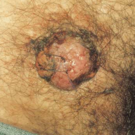
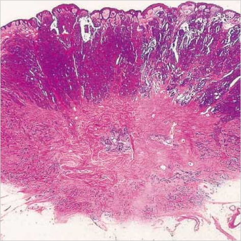
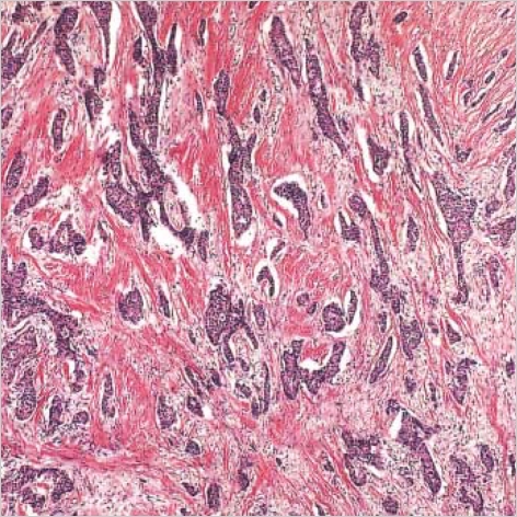
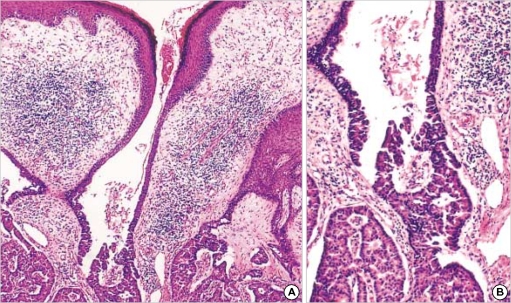
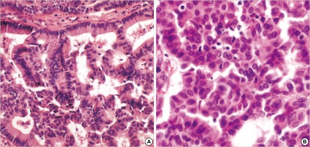
Syringocystadenocarcinoma papilliferum (SCACP) is a rare form of adenocarcinoma of the skin. This is the malignant counterpart of syringocystadenoma papilliferum (SCAP) and usually develops on the scalp in a long-standing lesion identified clinically as SCAP. We describe a 65-yr-old Korean man with a nodule on the right supra-pubic area with a 2-yr duration. Histologically this tumor had a similar overall configuration as in SCAP, but the tumor was asymmetric and poorly circumscribed, extending into the deep dermis and showed cytologic atypia. The tumor cells showed positive reaction to GCDFP-15, but negative reaction to CEA and HMFG-1. We established the diagnosis of SCACP in the patient, and a wide excision was performed to remove the tumor. The patient has been well without relapse or metastasis for 2 yr.
Figures
References
-
- Numata M, Hosoe S, Itoh N, Munakata Y, Hayashi S, Maruyama Y. Syringoadenocarcinoma papilliferum. J Cutan Pathol. 1985;12:3–7. - PubMed
-
- Requena L, Kiryu H, Ackerman AB. Ackerman's Histologic Diagnosis of Neoplastic Skin Disease: A Method by Pattern Analysis. Neoplasms with Apocrine Differentiation. Philadelphia, PA: Lippincott-Raven; 1998. pp. 665–675.
-
- Dissanayake RV, Salm R. Sweat-gland carcinomas. Prognosis related to histological type. Histopathology. 1980;4:445–466. - PubMed
-
- Seco Navedo MA, Fresno Forcelledo M, Orduna Domingo A, Junco Petrement P, Soler Sanchez T. Syringocystadenoma papilliferum with malignant evolution: presentation of a case. Ann Dermatol Venereol. 1982;109:685–689. - PubMed
-
- Bonadi R, Urso C. Syringocystadenocarcinoma papilliferum. Histopathology. 1996;28:475–477. - PubMed
Publication types
MeSH terms
Substances
LinkOut - more resources
Full Text Sources
