

Reusable, reversibly sealable parylene membranes for cell and protein patterning
- PMID: 17729252
- PMCID: PMC2841044
- DOI: 10.1002/jbm.a.31281
Reusable, reversibly sealable parylene membranes for cell and protein patterning
Abstract
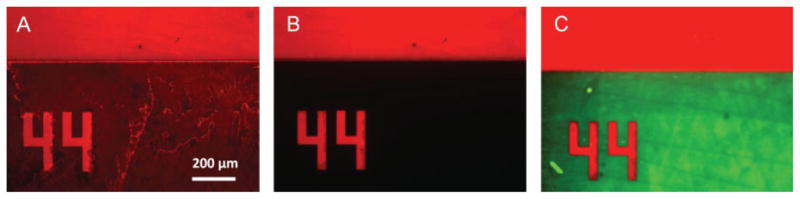
The patterned deposition of cells and biomolecules on surfaces is a potentially useful tool for in vitro diagnostics, high-throughput screening, and tissue engineering. Here, we describe an inexpensive and potentially widely applicable micropatterning technique that uses reversible sealing of microfabricated parylene-C stencils on surfaces to enable surface patterning. Using these stencils it is possible to generate micropatterns and copatterns of proteins and cells, including NIH-3T3 fibroblasts, hepatocytes and embryonic stem cells. After patterning, the stencils can be removed from the surface, plasma treated to remove adsorbed proteins, and reused. A variety of hydrophobic surfaces including PDMS, polystyrene and acrylated glass were patterned using this approach. Furthermore, we demonstrated the reusability and mechanical integrity of the parylene membrane for at least 10 consecutive patterning processes. These parylene-C stencils are potentially scalable commercially and easily accessible for many biological and biomedical applications.
Copyright 2007 Wiley Periodicals, Inc.
Figures


Similar articles
-
Generation of static and dynamic patterned co-cultures using microfabricated parylene-C stencils.Lab Chip. 2007 Oct;7(10):1272-9. doi: 10.1039/b706081e. Epub 2007 Jul 25. Lab Chip. 2007. PMID: 17896010
-
Microfabricated multilayer parylene-C stencils for the generation of patterned dynamic co-cultures.J Biomed Mater Res A. 2008 Jul;86(1):278-88. doi: 10.1002/jbm.a.32030. J Biomed Mater Res A. 2008. PMID: 18442109
-
Cell and protein compatibility of parylene-C surfaces.Langmuir. 2007 Nov 6;23(23):11718-25. doi: 10.1021/la7017049. Epub 2007 Oct 4. Langmuir. 2007. PMID: 17915896
-
Layered patterning of hepatocytes in co-culture systems using microfabricated stencils.Biotechniques. 2010 Jan;48(1):47-52. doi: 10.2144/000113317. Biotechniques. 2010. PMID: 20078427 Free PMC article.
-
Serum protein layers on parylene-C and silicon oxide: effect on cell adhesion.Colloids Surf B Biointerfaces. 2015 Feb 1;126:169-77. doi: 10.1016/j.colsurfb.2014.12.020. Epub 2014 Dec 16. Colloids Surf B Biointerfaces. 2015. PMID: 25555155 Free PMC article.
Cited by
-
A rapid co-culture stamping device for studying intercellular communication.Sci Rep. 2016 Oct 18;6:35618. doi: 10.1038/srep35618. Sci Rep. 2016. PMID: 27752145 Free PMC article.
-
Robust and Gradient Thickness Porous Membranes for In Vitro Modeling of Physiological Barriers.Adv Mater Technol. 2020 Dec;5(12):2000474. doi: 10.1002/admt.202000474. Epub 2020 Nov 9. Adv Mater Technol. 2020. PMID: 33709013 Free PMC article.
-
Established and novel methods of interrogating two-dimensional cell migration.Integr Biol (Camb). 2012 Nov;4(11):1338-50. doi: 10.1039/c2ib20154b. Integr Biol (Camb). 2012. PMID: 23038152 Free PMC article. Review.
-
A micropore array-based solid lift-off method for highly efficient and controllable cell alignment and spreading.Microsyst Nanoeng. 2020 Sep 7;6:86. doi: 10.1038/s41378-020-00191-5. eCollection 2020. Microsyst Nanoeng. 2020. PMID: 34567696 Free PMC article.
-
Cell patterning on photolithographically defined parylene-C: SiO2 substrates.J Vis Exp. 2014 Mar 7;(85):50929. doi: 10.3791/50929. J Vis Exp. 2014. PMID: 24637580 Free PMC article.
References
-
- Folch A, Toner M. Microengineering of cellular interactions. Annu Rev Biomed Eng. 2000;2:227–256. - PubMed
-
- Sims CE, Allbritton NL. Analysis of single mammalian cells on-chip. Lab Chip. 2007;7:423–440. - PubMed
-
- James CD, Davis R, Meyer M, Turner A, Turner S, Withers G, Kam L, Banker G, Craighead H, Issacson M, Turner J, Shain W. Aligned microcontact printing of micrometer-scale poly-L-lysine structures for controlled growth of cultured neurons on planar microelectrode arrays. IEEE Trans Biomed Eng. 2000;47:17–21. - PubMed
-
- Lahiri J, Ostuni E, Whitesides GM. Patterning ligands on reactive SAMs by microcontact printing. Langmuir. 1999;15:2055–2060.
Publication types
MeSH terms
Substances
Grants and funding
LinkOut - more resources
Full Text Sources
Other Literature Sources
