Review
. 2007 Nov;9(11):1883-98.
doi: 10.1089/ars.2007.1809.

Molecular targeting of proteins by L-homocysteine: mechanistic implications for vascular disease

Affiliations
Review

Molecular targeting of proteins by L-homocysteine: mechanistic implications for vascular disease

Alla V Glushchenko et al. Antioxid Redox Signal. 2007 Nov.

Abstract

Hyperhomocysteinemia is an independent risk factor for cardiovascular disease, complications of pregnancy, cognitive impairment, and osteoporosis. That elevated homocysteine leads to vascular dysfunction may be the linking factor between these apparently unrelated pathologies. Although a growing body of evidence suggests that homocysteine plays a causal role in atherogenesis, specific mechanisms to explain the underlying pathogenesis have remained elusive. This review focuses on chemistry unique to the homocysteine molecule to explain its inherent cytotoxicity. Thus, the high pKa of the sulfhydryl group (pKa, 10.0) of homocysteine underlies its ability to form stable disulfide bonds with protein cysteine residues, and in the process, alters or impairs the function of the protein. Studies in this laboratory have identified albumin, fibronectin, transthyretin, and metallothionein as targets for homocysteinylation. In the case of albumin, the mechanism of targeting has been elucidated. Homocysteinylation of the cysteine residues of fibronectin impairs its ability to bind to fibrin. Homocysteinylation of the cysteine residues of metallothionein disrupts zinc binding by the protein and abrogates inherent superoxide dismutase activity. Thus, S-homocysteinylation of protein cysteine residues may explain mechanistically the cytotoxicity of elevated L-homocysteine.


Figures

FIG. 1
FIG. 1. Buried and exposed conformations of albumin-Cys34 thiolate anion
Cys34 exists in buried and exposed conformations, as proposed by Christodoulou et al. (16). When albumin-Cys34 thiolate anion (exposed) attacks homocystine (Hcy-S-S-Hcy), the homocysteinylated product is stabilized in the exposed conformation. Modified from Christodoulou et al. (16). Reprinted by permission from Sengupta et al. (112).
FIG. 2
FIG. 2. Model for the formation of homocystine, homocysteine-cysteine mixed disulfide (HCMD), cystine, and albumin-bound homocysteine in circulation
Free reduced cysteine (Cys-SH) entering circulation is autooxidized by ceruloplasmin to cystine (Cys-S-S-Cys). Albumin thiolate anion enters the circulation and reacts with cystine to form albumin-Cys34-S-S-cysteine and cysteine thiolate anion. Free reduced homocysteine (Hcy-SH) entering circulation then attacks albumin-Cys34-S-S-cysteine to form HCMD (Hcy-S-S-Cys) and albumin thiolate anion. Albumin thiolate anion then reacts with HCMD, preferentially forming albumin-Cys34-S-S-homocysteine and cysteine thiolate anion. A small amount of homocysteine is also autooxidized to homocystine (Hcy-S-S-Hcy) by the copper attached to His3 of albumin. Albumin thiolate anion also reacts with homocystine to form albumin-Cys34-S-S-homocysteine. Thick arrows, The major reactions. Reprinted by permission from Sengupta et al. (113).
FIG. 3
FIG. 3. Phosphorimage analysis of the binding of 35S-d,l-homocysteine to albumin and transthyretin
Human plasma from a healthy donor (lanes 1 and 3) and purified transthyretin (lanes 2 and 4) were incubated with 35S- d,l-homocysteine for 5 h at 37°C. The samples, before (lanes 1 and 2) and after (lanes 3 and 4) treatment with 2-mercaptoethanol (BME), were subjected to SDS-PAGE followed by phosphorimage analysis. Reprinted by permission from Lim et al. (67).
FIG. 4
FIG. 4. Binding of l-homocysteine to transthyretin (in vivo)
Deconvoluted electrospray ionization mass spectra of transthyretin, immunoprecipitated and HPLC-purified, from (A) the plasma of a patient with end-stage renal disease (20.7 µM total plasma homocysteine) and (B) from the plasma of a patient with homocystinuria (434 µM total plasma homocysteine) are shown. Reprinted by permission from Lim et al. (67).
FIG. 5
FIG. 5. The binding of homocysteine and cysteine to plasma proteins and purified fibronectin
35S-d,l-Homocysteine (A) and 35S-l-cysteine (500 µM final concentration) (B) were incubated with 50% human plasma in 0.05 M TES buffer (pH 7.4) and with human fibronectin (1 mg/ml in 0.05 M TES buffer, pH 7.4) for 5 h at 37°C. After the incubation, protein was precipitated with 1.5 M perchloric acid and solubilized with SDS-PAGE sample buffer. Aliquots were subjected to analysis by SDS-PAGE. The gels were stained with Coomassie blue, dried, and subjected to phosphorimaging. (A) Binding of 35S-d,l-homocysteine. Lanes are as follows: 1 and 3, human plasma; 2 and 4, human plasma fibronectin. Lanes 1 and 2 are without reduction, and lanes 3 and 4 are with reduction by 2-mercaptoethanol. (B) Binding of 35S-l-cysteine. Lanes are as follows: 1, human plasma; 2, human plasma fibronectin. Reprinted by permission from Majors et al. (73).
FIG. 6
FIG. 6. Identification of 35S-d,l-homocysteinylated-metallothionein (MT) in human aortic endothelial cells (HAECs)
(A) Western blot of HAEC lysate probed with anti-MT antibody in the absence (NR, nonreducing) and presence (R, reducing) of É-mercaptoethanol (BME). (B) Phosphorimage of the same Western blot depicted in (A), demonstrating 35S-homocysteinylated proteins. Several bands including the one corresponding to MT are present in the NR lane but not in the R lane. Reprinted with permission from Barbato et al. (3).
FIG. 7
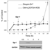
FIG. 7. Intracellular free Zn2+, ROS, and Egr-1 protein expression in HAECs as a function of time exposed to l-homocysteine
Solid line, The percentage change in intracellular free Zn2+ in HAECs incubated with 50 µM l-homocysteine. Dashed line, The percentage change in ROS as measured with CM-H2DCFDA–loaded HAECs incubated with 50 µM l-homocysteine. The inset depicts a Western blot probed with anti-Egr-1 and GAPDH antibodies at 0.5-, 1-, 2-, and 4-h points. Arrow, Egr-1 protein expression. Error bars represent ±SD. *Statistical significance compared with Zinquin-loaded cells at the zero time point (p < 0.01). †Statistical significance compared with CM-H2DCFDA–loaded cells at the zero time point (p < 0.01). Reprinted with permission from Barbato et al. (3).
FIG. 8
FIG. 8. Direct frontal attack of homocysteine (Hcy) on the ER
Molecular targeting of the ER plasma membrane, ER chaperones (e.g., GRP78), ER processing machinery, or client proteins by homocysteine may induce ER stress. ER stress results in the activation of the intracellular signaling pathway, called the unfolded protein response (UPR). Genes encoding ER chaperones and processing enzymes become transcriptionally upregulated. In eukaryotes, the UPR is regulated by the proximal sensors PKR-like ER kinase (PERK), activating transcription factor 6 (ATF6), and type-I ER transmembrane protein kinase (IRE1). Activation of these sensors leads to increased expression of ER-resident chaperones (GRP78) and the other UPR proteins. ER stress leads to a decrease in protein synthesis and translation through PERK. In late-stage ER stress, PERK, ATF6, and IRE1 upregulate CHOP (C/EBP homologous protein), promoting cell death (apoptosis). SREBP (sterol regulatory element–binding protein) upregulates lipid synthesis. Prolonged UPR leads to Ca2+ release from the ER, causing production of ROS, which may lead to activation of NF-κB. Activation of these downstream pathways after ER stress can have direct effects on atherosclerotic lesion development and thrombogenicity.
FIG. 9
FIG. 9. Determination of the macroscopic sulfhydryl group pKa values for l-homocysteine (left) and l-cysteine (right)
Absorbance of the thiolate anion (RS) was measured at 240 nm at the indicated pH in physiologic saline and buffer. The final concentration of l-homocysteine and l-cysteine was 250 µM. (Jacobsen, Catanescu, and Abramczyk, unpublished observations).

Similar articles

Cited by

References

    1. Austin RC, Lentz SR, Werstuck GH. Role of hyperhomocysteinemia in endothelial dysfunction and atherothrombotic disease. Cell Death Differ. 2004;11 suppl 1:S56–S64. - PubMed
    1. Banhegyi G, Csala M, Nagy G, Sorrentino V, Fulceri R, Benedetti A. Evidence for the transport of glutathione through ryanodine receptor channel type 1. Biochem J. 2003;376:807–812. - PMC - PubMed
    1. Barbato JC, Catanescu O, Murray K, Dibello PM, Jacobsen DW. Targeting of metallothionein by L-homocysteine: a novel mechanism for disruption of zinc and redox homeostasis. Arterioscler Thromb Vasc Biol. 2007;27:49–54. - PMC - PubMed
    1. Beckmann AM, Wilce PA. Egr transcription factors in the nervous system. Neurochem Int. 1997;31:477–510. discussion 517–476. - PubMed
    1. Benson MD. Amyloidosis. In: Scriver CR, Beaudet AL, Sly WS, Valle D, editors. The metabolic and molecular bases of inherited disease. New York: McGraw-Hill Medical Publishing Division; 2001. pp. 5345–5378.