

Mast cells in vulnerable atherosclerotic plaques--a view to a kill
- PMID: 17760836
- PMCID: PMC3823253
- DOI: 10.1111/j.1582-4934.2007.00052.x
Mast cells in vulnerable atherosclerotic plaques--a view to a kill
Abstract
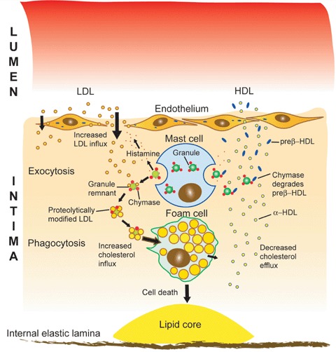
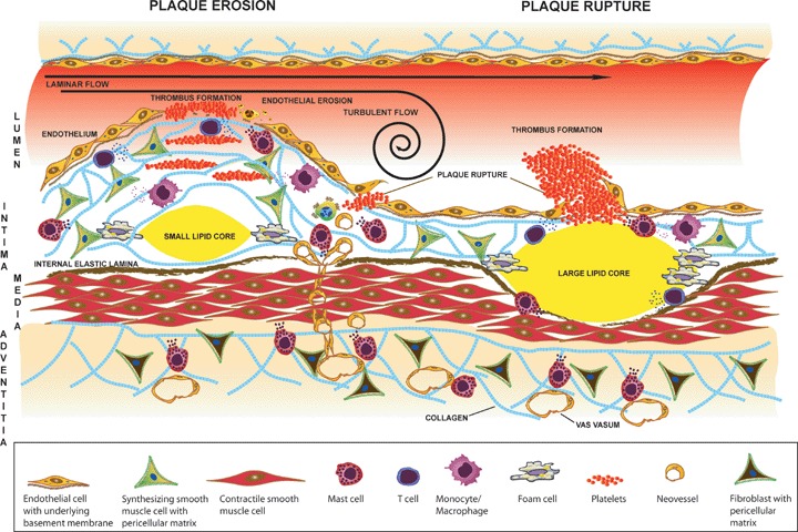
The aim of the present review is to discuss the participation of mast cells in the pathogenesis of erosion and rupture of atherosclerotic plaques, the major causes behind acute coronary syndromes and myocardial infarction. We present ex vivo observations describing mast cells and their activation in human atherosclerotic plaques and discuss in vitro and in vivo data showing that mast cells are potential regulators of inflammation, immunity and adverse remodeling, including matrix remodeling and cell death. Furthermore, we focus on studies that have been performed with human tissues and human mast cells, but when appropriate, we also discuss observations made in animal models. Finally, we present potential pharmacological means to modulate mast cell responses in the arterial vessel walls.
Figures
Similar articles
-
Mast cells and degradation of pericellular and extracellular matrices: potential contributions to erosion, rupture and intraplaque haemorrhage of atherosclerotic plaques.Biochem Soc Trans. 2007 Nov;35(Pt 5):857-61. doi: 10.1042/BST0350857. Biochem Soc Trans. 2007. PMID: 17956232 Review.
-
The role of mast cells in atherosclerosis.Hamostaseologie. 2015;35(2):113-20. doi: 10.5482/HAMO-14-08-0034. Epub 2014 Nov 7. Hamostaseologie. 2015. PMID: 25377048 Review.
-
Chlamydia pneumoniae and atherosclerosis: the role of mast cells.J Biol Regul Homeost Agents. 2009 Apr-Jun;23(2):65-9. J Biol Regul Homeost Agents. 2009. PMID: 19589286 Review.
-
Mast cells in vulnerable coronary plaques: potential mechanisms linking mast cell activation to plaque erosion and rupture.Curr Opin Lipidol. 2004 Oct;15(5):567-73. doi: 10.1097/00041433-200410000-00011. Curr Opin Lipidol. 2004. PMID: 15361793 Review.
-
[Immunohistochemical study of the role of mast cells and macrophages in the process of angiogenesis in the atherosclerotic plaques in patients with metabolic syndrome].Arkh Patol. 2016 Mar-Apr;78(2):19-28. doi: 10.17116/patol201678219-28. Arkh Patol. 2016. PMID: 27070771 Russian.
Cited by
-
Glucagon effects on 3H-histamine uptake by the isolated guinea-pig heart during anaphylaxis.Biomed Res Int. 2014;2014:782709. doi: 10.1155/2014/782709. Epub 2014 May 11. Biomed Res Int. 2014. PMID: 24895609 Free PMC article.
-
New Insights into the Role of Inflammation in the Pathogenesis of Atherosclerosis.Int J Mol Sci. 2017 Sep 22;18(10):2034. doi: 10.3390/ijms18102034. Int J Mol Sci. 2017. PMID: 28937652 Free PMC article. Review.
-
Micro- and Macrovascular Effects of Inflammation in Peripheral Artery Disease-Pathophysiology and Translational Therapeutic Approaches.Biomedicines. 2023 Aug 17;11(8):2284. doi: 10.3390/biomedicines11082284. Biomedicines. 2023. PMID: 37626780 Free PMC article. Review.
-
Mast cell derived carboxypeptidase A3 is decreased among patients with advanced coronary artery disease.Cardiol J. 2019;26(6):680-686. doi: 10.5603/CJ.a2018.0018. Epub 2018 Mar 7. Cardiol J. 2019. PMID: 29512095 Free PMC article.
-
Elevated Adiponectin Levels Suppress Perivascular and Aortic Inflammation and Prevent AngII-induced Advanced Abdominal Aortic Aneurysms.Sci Rep. 2016 Sep 23;6:31414. doi: 10.1038/srep31414. Sci Rep. 2016. PMID: 27659201 Free PMC article.
References
-
- Kirshenbaum AS, Kessler SW, Goff JP, Metcalfe DD. Demonstration of the origin of human mast cells from CD34+ bone marrow progenitor cells. J Immunol. 1991;146:1410–5. - PubMed
-
- Rottem M, Okada T, Goff JP, Metcalfe DD. Mast cells cultured from the peripheral blood of normal donors and patients with mastocytosis originate from a CD34+/Fc epsilon RI- cell population. Blood. 1994;84:2489–96. - PubMed
-
- Kirshenbaum AS, Goff JP, Semere T, Foster B, Scott LM, Metcalfe DD. Demonstration that human mast cells arise from a progenitor cell population that is CD34(+), c-kit(+), and expresses aminopeptidase N (CD13) Blood. 1999;94:2333–42. - PubMed
-
- Boyce JA, Mellor EA, Perkins B, Lim YC, Luscinskas FW. Human mast cell progenitors use alpha4-integrin, VCAM-1, and PSGL-1 E-selectin for adhesive interactions with human vascular endothelium under flow conditions. Blood. 2002;99:2890–6. - PubMed
Publication types
MeSH terms
LinkOut - more resources
Full Text Sources
Other Literature Sources
Medical
