. 2007 Jul-Aug;11(4):764-75.
doi: 10.1111/j.1582-4934.2007.00066.x.

Close relation of arterial ICC-like cells to the contractile phenotype of vascular smooth muscle cell

Affiliations

Close relation of arterial ICC-like cells to the contractile phenotype of vascular smooth muscle cell

Vladimír Pucovský et al. J Cell Mol Med. 2007 Jul-Aug.

Abstract

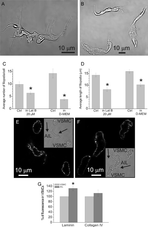
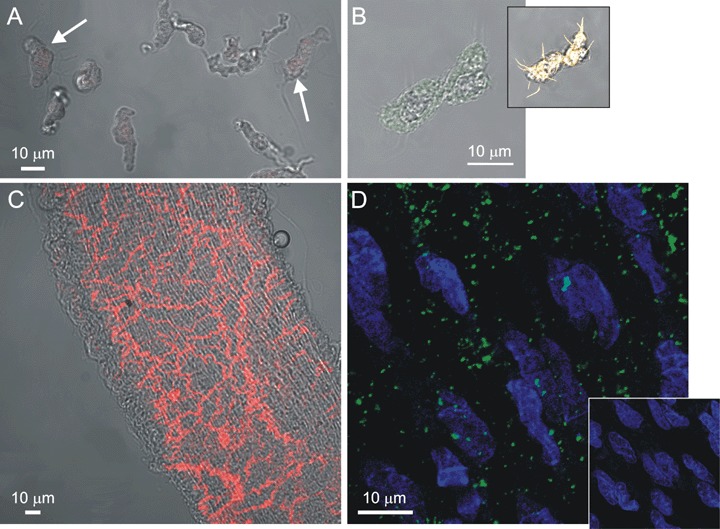
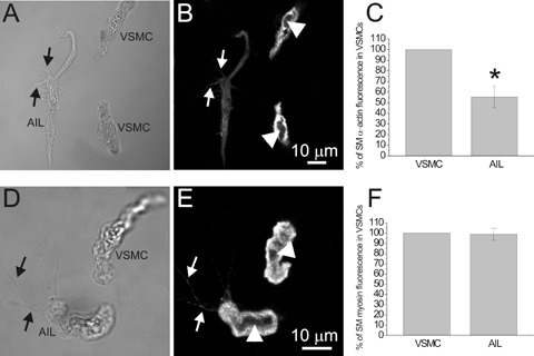
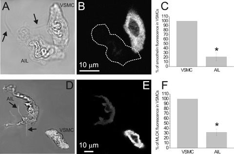
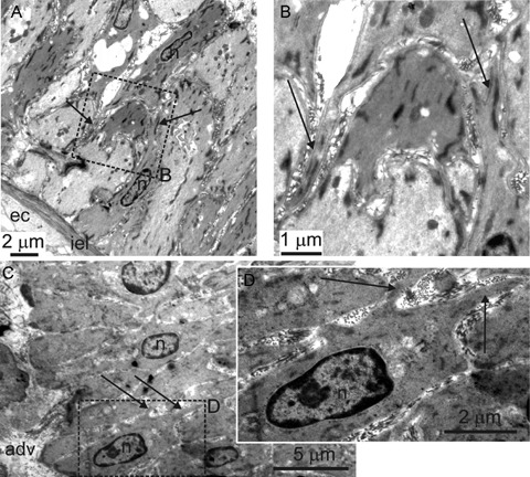
This work aimed to establish the lineage of cells similar to the interstitial cells of Cajal (ICC), the arterial ICC-like (AIL) cells, which have recently been described in resistance arteries, and to study their location in the artery wall. Segments of guinea-pig mesenteric arteries and single AIL cells freshly isolated from them were used. Confocal imaging of immunostained cells or segments and electron microscopy of artery segments were used to test for the presence and cellular localization of selected markers, and to localize AIL cells in intact artery segments. AIL cells were negative for PGP9.5, a neural marker, and for von Willebrand factor (vWF), an endothelial cell marker. They were positive for smooth muscle alpha-actin and smooth muscle myosin heavy chain (SM-MHC), but expressed only a small amount of smoothelin, a marker of contractile smooth muscle cells (SMC), and of myosin light chain kinase (MLCK), a critical enzyme in the regulation of smooth muscle contraction. Cell isolation in the presence of latrunculin B, an actin polymerization inhibitor, did not cause the disappearance of AIL cells from cell suspension. The fluorescence of basal lamina protein collagen IV was comparable between the AIL cells and the vascular SMCs and the fluorescence of laminin was higher in AIL cells compared to vascular SMCs. Moreover, cells with thin processes were found in the tunica media of small resistance arteries using transmission electron microscopy. The results suggest that AIL cells are immature or phenotypically modulated vascular SMCs constitutively present in resistance arteries.


Figures

4
4
(A) Transmitted light image of an AIL cell isolated using the usual procedure. (B) Transmitted light image of three AIL cells isolated in the presence of 20 μM latrunculin B. (C) The average number of filopodia per cell was decreased from 9.9 ± 1.0 per cell, n = 25 in control (Ctrl) to 6.4 ± 0.5 per cell, n = 39 in latrunculin B (Lat B), P = 0.0006, significantly different. Cell isolation in Dulbecco's Modified Eagle's Medium (D-MEM) also decreased it from 14.3 ± 1.8 per cell, n = 10 in control to 3.8 ± 0.4 per cell, n = 14, P = 0.000002, significantly different. (D) The average length of filopodia was decreased from 14.4 ± 0.8 μm, n = 99 in control to 8.2 ± 0.8 μm, n = 55 in latrunculin B, P = 0.000003, significantly different, and from 16.0 ± 0.7 μm, n = 143 in control to 10.3 ± 0.7 μm in D-MEM, n = 53, P = 0.00003, significantly different. (E) Image of laminin fluorescence in an AIL cell and two VSMCs obtained by confocal microscopy. Inset shows the same cells in transmitted light. Arrows point to some of the filopodia. (F) Same as in (E), but staining for collagen IV is shown. (G) Laminin fluorescence of AIL cells was 131.9 ± 9.6% of that in VSMCs (n = 8 cell pairs, P = 0.013, significantly different) and collagen IV fluorescence was 113.3 ± 12.1% of that in VSMCs (n = 9 cell pairs, P = 0.305). Omitting primary antibodies produced virtually no fluorescence in both cases (n = 13 and n = 12, respectively; not shown). * Statistically significant difference to control.
1
1
Fluorescent staining for PGP9.5 and vWF. (A) A combined image of transmitted light (shades of grey) and PGP9.5 fluorescence (red) of VSMCs and AIL cells (arrows) obtained by confocal microscopy. Neither AIL cells nor VSMCs stained positive for PGP9.5 (n = 24 cells). (B) A combined image of transmitted light (shades of grey) and vWF fluorescence (green) of an AIL cell, showing only weak non-specific staining (n = 6). Inset: Same cell stained with BODIPY 558/568 phalloidin (yellow) revealed the presence of otherwise faintly visible filopodia, confirming the identity of an AIL cell. (C) Same as in (A), but a segment of mesenteric artery was used, showing the presence of nerve fibers (positive control, n = 5). (D) A fluorescent image of an endothelial layer in mesenteric artery segment immunostained for vWF and stained with SYTO 40 for nucleic acids. Nuclei of endothelial cells can be seen in blue. Green colour shows vWF staining of granular appearance, due to its storage in the Weibel-Palade bodies of endothelial cells (n = 4). Inset: As for the main panel, but the primary antibodies to vWF were omitted.
2
2
Fluorescent staining for SMA and SM-MHC. (A) Transmitted light image of an AIL cell and two VSMCs and (B) an image of SMA fluorescence in the same cells, obtained by confocal microscopy. (C) SMαA fluorescence of AIL cells was 55.5 ± 10.2% of that in VSMCs (n = 6 cell pairs, P = 0.0074, significantly different). Omitting primary antibodies (n = 9) produced in average 1.8% of the original fluorescence intensity (n = 6; not shown). (D) and (E) As for (A) and (B), but staining for SM-MHC is shown. (F) SM-MHC fluorescence of AIL cells was 98.9 ± 5.8% of that in VSMCs (n = 7 cell pairs, P = 0.852). Omitting primary antibodies (n = 10) produced an average of less than 0.1% of the original fluorescence intensity (n = 7; not shown). Arrows point to filopodia of AIL cells. In both cases, fluorescence was found throughout the cytoplasm of VSMCs and AIL cells, except for the centre, occupied by nucleus (arrowheads). * Statistically significant.
3
3
Fluorescent staining for smoothelin and MLCK. (A) Transmitted light image of an AIL cell and a VSMC and (B) an image of smoothelin fluorescence in the same cells, obtained by confocal microscopy. (C) Smoothelin fluorescence of AIL cells was 21.4 ± 8.7% of that in VSMCs (n = 10 cell pairs from three animals, P = 0.00001, statistically significant). Omitting primary antibodies (n = 29 cells from five animals) produced an average of 2.6% of the original fluorescence intensity (n = 10; not shown). (D) Transmitted light image of an AIL cell and a VSMC combined with the image of BODIPY phalloidin fluorescence of the bottom plane (in white; to enhance the visibility of filopodia). (E) As for (B), but staining for MLCK is shown. (F) MLCK fluorescence of AIL cells was 32.1 ± 6.6% of that in VSMCs (n = 9 cell pairs, P = 0.00001, statistically significant). Omitting primary antibodies (n = 7) produced an average of 8.7% of the original fluorescence intensity (n = 9; not shown). The fluorescence of both markers was localized throughout the cytoplasm of VSMCs and AIL cells, except for the nucleus. Arrows point to filopodia of AIL cells. * Statistically significant.
5
5
Cells with thin processes in intact mesenteric arteries. Transmission electron micrographs of an oblique section through mesenteric artery. Rectangles in (A) and (C) delineated by dashed line indicate areas which are shown enlarged in (B) and (D), respectively. Adv – adventitia, ec – endothelial cell, iel – internal elastic lamina, n – nucleus. Arrows indicate processes. The cells with thin processes can be seen in the tunica media.

References

    1. Campbell GR, Campbell JH, Manderson JA, Horrigan S, Rennick RE. Arterial smooth muscle: a multifunctional mesenchymal cell. Arch Pathol Lab Med. 1988;112:977–86. - PubMed
    1. Chamley-Campbell JH, Campbell GR. Wha? controls smooth muscle phenotype? Atherosclerosis. 1981;40:347–57. - PubMed
    1. Shanahan CM, Weissberg PL. Smooth muscle cell heterogeneity. Patterns of gene expression in vascular smooth muscle cells in vitro and in vivo. Arterioscler Thromb Vasc Biol. 1998;18:333–8. - PubMed
    1. Sartore S, Chiavegato A, Faggin E, Franch R, Puato M, Ausoni S, Pauletto P. Contribution of adventitial fibroblasts to neointima formation and vascular remodeling. From innocent bystander to active participant. Circ Res. 2001;89:1111–21. - PubMed
    1. Halayko AJ, Solway J. Plasticity in skeletal, cardiac, and smooth muscle. Invited review: molecular mechanisms of phenotypic plasticity in smooth muscle cells. J Appl Physiol. 2001;90:358–68. - PubMed

Publication types

MeSH terms