. 2007 Jul-Aug;11(4):810-25.
doi: 10.1111/j.1582-4934.2007.00074.x.

Studies on brain volume, Alzheimer-related proteins and cytokines in mice with chronic overexpression of IL-1 receptor antagonist

Affiliations

Studies on brain volume, Alzheimer-related proteins and cytokines in mice with chronic overexpression of IL-1 receptor antagonist

M Oprica et al. J Cell Mol Med. 2007 Jul-Aug.

Abstract

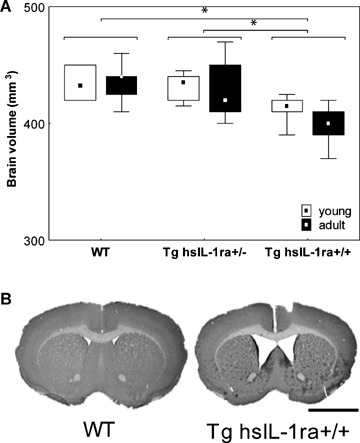
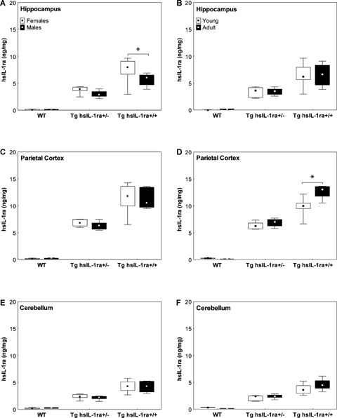
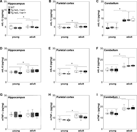
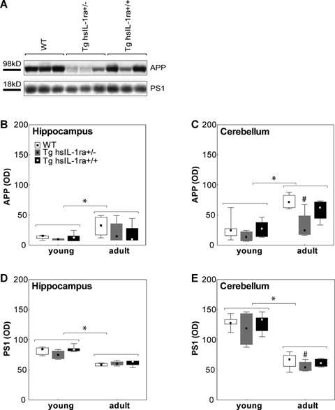
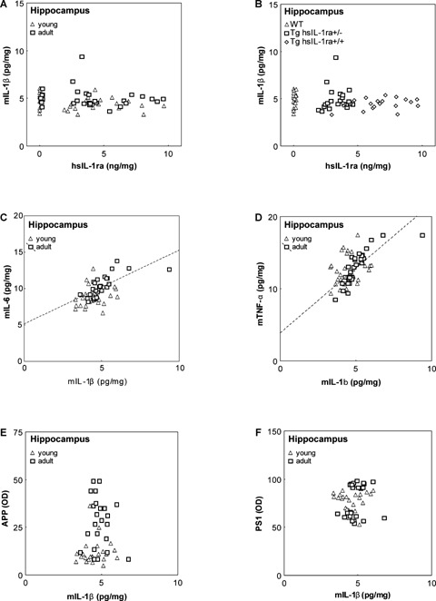
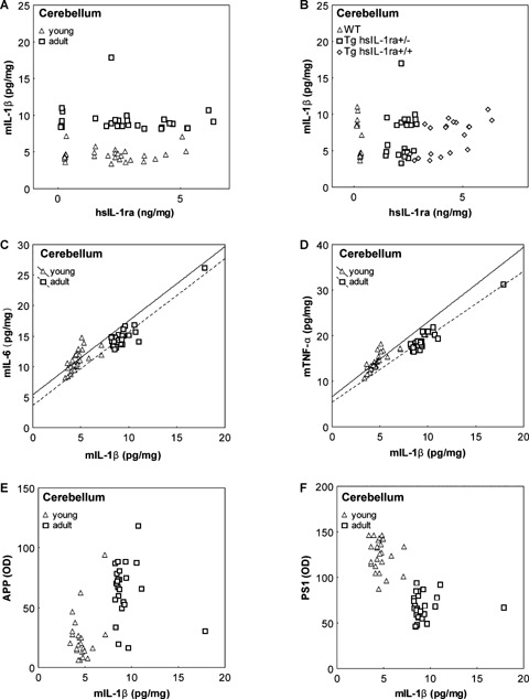
Inflammation is associated with both acute and chronic neurological disorders, including stroke and Alzheimer's disease (AD). Cytokines such as interleukin (IL)-1 have several activities in the brain both under physiological and pathophysiological conditions. The objective of this study was to evaluate consequences of the central blockade of IL-1 transmission in a previously developed transgenic mouse strain with brain-directed overexpression of human soluble IL-1 receptor antagonist (Tg hsIL-1ra). Effects on brain morphology and brain levels of the AD-related proteins beta-amyloid precursor protein (APP) and presenilin 1(PS1), as well as the levels of IL-1beta, IL-6 and tumour necrosis factor-alpha (TNF-alpha) were analysed in homozygotic and heterozygotic mice and wild type (WT) controls, of both genders and of young (30-40 days) and adult (13-14 months) age. A marked reduction in brain volume was observed in transgenic mice as determined by volumetry. Western blot analysis showed higher levels of APP, but lower levels of PS1, in adult animals than in young ones. In the cerebellum, heterozygotic (Tg hsIL-1ra(+/-)) mice had lower levels of APP and PS1 than WT mice. With one exception, there were no genotypic differences in the levels of IL-1beta, IL-6 and TNF-alpha. The cytokine levels were generally higher in adult than in young mice. In conclusion, the chronic blockade of IL-1 signalling in the brain was associated with an atrophic phenotype of the brain, and with modified levels of APP and PS1. Brain-directed overexpression of hsIL-1ra was not followed by major compensatory changes in the levels of pro-inflammatory cytokines.


Figures

1
1
Total brain volume (A) in mice with transgenic expression of human soluble interleukin-1 receptor antagonist (hsIL-1ra) with homozygotic (Tg hsIL-1ra+/+) and heterozygotic (Tg hsIL-1ra+/−) genotype, and of wild type (WT) mice, of young (1month) and adult (13–14 months) age. Tg hsIL-1ra+/+ mice have a smaller brain volume than Tg IL-1ra+/− mice (*P < 0.001) and WT mice (*P < 0.001), respectively (A). B shows cresyl violet-stained sections of the brain at bregma +0.6 mm, from two adult mice, WT and homozygotic (Tg hsIL-1ra+/+), respectively, illustrating the reduced brain volume and enlarged ventricles. Scale bar = 2.5 mm.
2
2
The levels of human interleukin-1 receptor antagonist (hIL-1ra) in hippocampus (A, B), parietal cortex (C, D) and cerebellum (E, F) of male and female mice with transgenic expression of human soluble IL-1ra (hsIL-1ra) with homozygotic (Tg hsIL-1ra+/+) and heterozygotic (Tg hsIL-1ra+/−) genotype, and of wild type (WT) mice. The levels are gene dosage-dependent in all the three brain regions (A–F; statistical data given in Results). In the hippocampus, female mice have higher levels than male mice (*P < 0.001) (A). An age-dependent increase is seen in the parietal cortex of homozygotic mice (*P < 0.001) (D).
3
3
The levels of mouse interleukin-1β (mIL-1β) (A-C), IL-6 (D-F) and TNF-α (G-I) in the hippocampus (A, D and G), parietal cortex (B, E and H) and cerebellum (C, F and I) of mice with transgenic expression of human soluble interleukin- 1 receptor antagonist (hsIL-1ra) with homozygotic (Tg hsIL-1ra+/+) and heterozygotic (Tg hsIL-1ra+/−) genotype, and of wild type (WT) mice, of young (1 month) and adult (13–14 months) age. Adult mice have higher levels of mIL- 1β than the young mice in the hippocampus (*P < 0.05) (A), parietal cortex (*P < 0.05) (B) and cerebellum (*P < 0.001) (C). The levels of mIL-6 are higher in the adult than young mice in the hippocampus (*P < 0.001) (D), parietal cortex (*P < 0.001) (E) and cerebellum (*P < 0.001) (F). Adult mice have higher levels of mTNF-α levels than young mice in the parietal cortex (*P < 0.001) (H) and cerebellum (*P < 0.001) (I), but not in the hippocampus (G).
4
4
The levels of amyloid precursor protein (APP) (A–C) and presenilin-1 (PS1) (A, D and E) in the hippocampus (B and D) and cerebellum (A, C and E) of mice with transgenic expression of human soluble interleukin-1 receptor antagonist (hsIL-1ra) with homozygotic (Tg hsIL-1ra+/+) and heterozygotic (Tg hsIL-1ra+/−) genotype, and of wild type (WT) mice. Adult mice display higher levels of APP than young mice, both in the hippocampus (*P < 0.05) (B) and cerebellum (*P < 0.001) (C). In the male mice, APP levels are lower in Tg hsIL-1ra+/− mice than in WT mice (#P < 0.05) (A, C; adult mice). The above line in A shows a representative Western blot for APP, at the size of the full-length protein, in samples from the cerebellum of adult male mice. Regarding PS1 levels, young mice have higher levels than adult mice both in the hippocampus (*P < 0.001) (D) and cerebellum (*P < 0.001) (E). Tg hsIL-1ra+/− mice have lower levels of PS1 than adult WT in both males and females (#P < 0.05) (E; adult mice). The below line in A shows a representative Western blot for PS1, at the size of the full-length protein, in samples from the cerebellum of adult male mice.
5
5
Correlations between the levels of mouse interleukin-1β (mIL-1β) and human soluble interleukin-1 receptor antagonist (hsIL-1ra) (A, B), mouse interleukin-6 (mIL-6) (C), mouse tumour necrosis factor-α (mTNF-β) (D), amyloid precursor protein (APP) (E) and presenilin 1 (PS1) (F), respectively, in the hippocampus of mice with transgenic expression of hsIL-1ra with homozygotic (Tg hsIL-1ra+/+) and heterozygotic (Tg hsIL-1ra+/−) genotype, and of wild type (WT) mice, of young and adult age. No correlation was found between the levels of mIL-1β and hIL-1ra (A). Labelling of the individual animals according to genotype illustrates the different levels of hsIL-1ra (B). A positive correlation was found between the levels of mIL-1β and those of mIL-6 in adult mice (r = 0.70, P < 0.001) (C), and between the levels of mIL-1β and those of mTNF-α in adult mice (r = 0.81, P < 0.001) (D). The levels of APP (E) and PS1 (F) do not correlate with the levels of mIL-1β.
6
6
Correlations between the levels of mouse interleukin-1β (mIL-1β) and human soluble interleukin-1 receptor antagonist (hsIL-1ra) (A, B), mouse interleukin-6 (mIL-6) (C), mouse tumour necrosis factor-a (mTNF-α) (D), amyloid precursor protein (APP) (E) and presenilin 1 (PS1) (F), respectively, in the cerebellum of mice with transgenic expression of hsIL-1ra with homozygotic (Tg hsIL-1ra+/+) and heterozygotic (Tg hsIL-1ra+/−) genotype, and of wild type (WT) mice, of young and adult age. No correlation was found between the levels of mIL-1β and hsIL-1ra (A). Labelling of the individual animals according to genotype illustrates the different levels of hsIL-1ra (B). Adult mice have higher levels of mIL-1β than young mice of all genotypes (A, B). A positive correlation was found between the levels of mIL-1β and those of mIL-6 in both young (r = 0.66, P < 0.001), and adult (r = 0.92, P < 0.001) mice (C), and between the levels of mIL-1β and those of mTNF-α in both young (r = 0.78, P < 0.001), and adult (r = 0.95, P < 0.001) mice (D). APP (E) and PS1 (F) levels display a well-defined clustering pattern according to the age of the animals, but no correlation with mIL-1β levels.

References

    1. Rothwell NJ, Luheshi GN. Interleukin 1 in the brain: biology, pathology and therapeutic target. Trends Neurosci. 2000;23:618–25. - PubMed
    1. Minami M, Kuraishi Y, Yabuuchi K, Yamazaki A, Satoh M. Induction of interleukin-1β mRNA in rat brain after transient forebrain ischemia. J Neurochem. 1992;58:390–2. - PubMed
    1. Stroemer RP, Rothwell NJ. Exacerbation of ischemic brain damage by localized striatal injection of interleukin- 1β in the rat. J Cereb Blood Flow Metab. 1998;18:833–9. - PubMed
    1. Blume AJ, Vitek MP. Focusing on IL-1-promotion of β-amyloid precursor protein synthesis as an early event in Alzheimer's disease. Neurobiol Aging. 1989;10:406–8. - PubMed
    1. Giulian D, Young DG, Woodward J, Brown DC, Lachman LB. Interleukin-1 is an astroglial growth factor in the developing brain. J Neurosci. 1988;8:709–14. - PMC - PubMed

Publication types