

Sodium ascorbate induces apoptosis in neuroblastoma cell lines by interfering with iron uptake
- PMID: 17760959
- PMCID: PMC2000471
- DOI: 10.1186/1476-4598-6-55
Sodium ascorbate induces apoptosis in neuroblastoma cell lines by interfering with iron uptake
Abstract
Background: Neuroblastoma (NB) is an extra-cranial solid tumour of childhood. In spite of the good clinical response to first-line therapy, complete eradication of NB cells is rarely achieved. Thus, new therapeutic strategies are needed to eradicate surviving NB cells and prevent relapse. Sodium ascorbate has been recently reported to induce apoptosis of B16 melanoma cells through down-regulation of the transferrin receptor, CD71. Since NB and melanoma share the same embryologic neuroectodermal origin, we used different human NB cell lines to assess whether the same findings occurred.
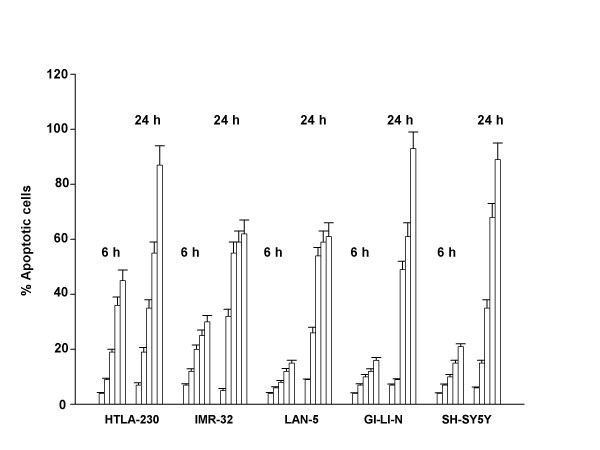
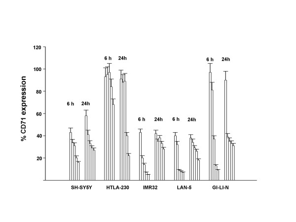
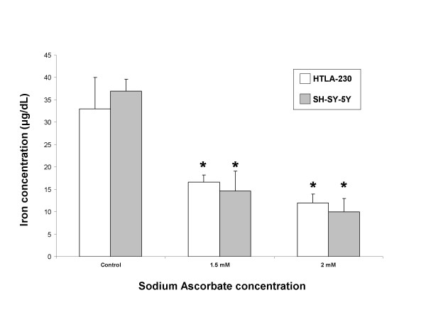
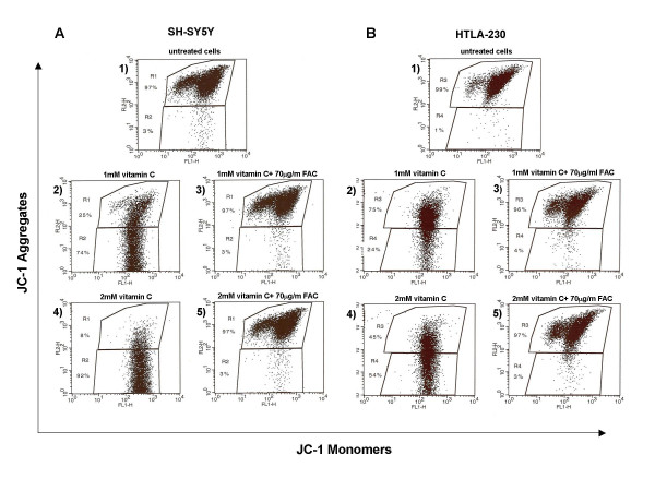
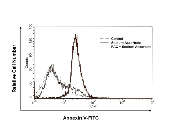
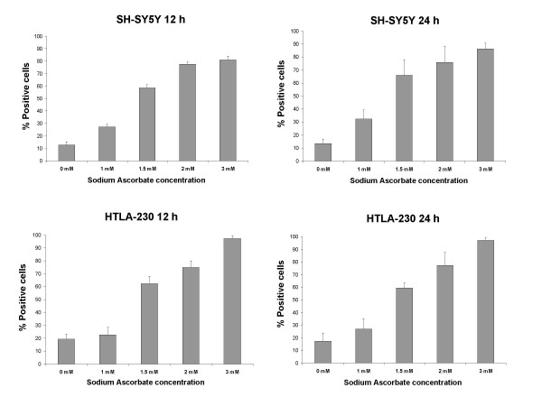
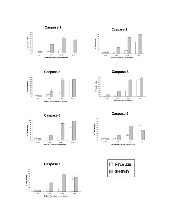
Results: We could observe dose- and time-dependent induction of apoptosis in all NB cell lines. Sodium ascorbate decreased the expression of CD71 and caused cell death within 24 h. An increase in the global and specific caspase activity took place, as well as an early loss of the mitochondrial transmembrane potential. Moreover, intracellular iron was significantly decreased after exposure to sodium ascorbate. Apoptotic markers were reverted when the cells were pretreated with the iron donor ferric ammonium citrate (FAC), further confirming that iron depletion is responsible for the ascorbate-induced cell death in NB cells.
Conclusion: Sodium ascorbate is highly toxic to neuroblastoma cell lines and the specific mechanism of vitamin C-induced apoptosis is due to a perturbation of intracellular iron levels ensuing TfR-downregulation.
Figures
Similar articles
-
Sodium ascorbate (vitamin C) induces apoptosis in melanoma cells via the down-regulation of transferrin receptor dependent iron uptake.J Cell Physiol. 2005 Jul;204(1):192-7. doi: 10.1002/jcp.20286. J Cell Physiol. 2005. PMID: 15672419
-
Sodium ascorbate inhibits growth via the induction of cell cycle arrest and apoptosis in human malignant melanoma A375.S2 cells.Melanoma Res. 2006 Dec;16(6):509-19. doi: 10.1097/01.cmr.0000232297.99160.9e. Melanoma Res. 2006. PMID: 17119452
-
Imatinib mesylate (STI571) interference with growth of neuroectodermal tumour cell lines does not critically involve c-Kit inhibition.Int J Mol Med. 2004 Sep;14(3):373-82. Int J Mol Med. 2004. PMID: 15289888
-
Ascorbic acid--important for iron metabolism.Folia Med (Plovdiv). 2008 Oct-Dec;50(4):11-6. Folia Med (Plovdiv). 2008. PMID: 19209525 Review.
-
Potential Treatment Options for Neuroblastoma with Polyphenols through Anti-Proliferative and Apoptotic Mechanisms.Biomolecules. 2023 Mar 20;13(3):563. doi: 10.3390/biom13030563. Biomolecules. 2023. PMID: 36979499 Free PMC article. Review.
Cited by
-
Role of Vitamin C in Targeting Cancer Stem Cells and Cellular Plasticity.Cancers (Basel). 2023 Nov 30;15(23):5657. doi: 10.3390/cancers15235657. Cancers (Basel). 2023. PMID: 38067361 Free PMC article. Review.
-
Inhibition of the SK-N-MC human neuroblastoma cell line in vivo and in vitro by a novel nutrient mixture.Oncol Rep. 2013 May;29(5):1714-20. doi: 10.3892/or.2013.2307. Epub 2013 Feb 27. Oncol Rep. 2013. PMID: 23446555 Free PMC article.
-
The Result of Vitamin C Treatment of Patients with Cancer: Conditions Influencing the Effectiveness.Int J Mol Sci. 2022 Apr 15;23(8):4380. doi: 10.3390/ijms23084380. Int J Mol Sci. 2022. PMID: 35457200 Free PMC article. Review.
-
A randomized phase II trial of best supportive care with or without hyperthermia and vitamin C for heavily pretreated, advanced, refractory non-small-cell lung cancer.J Adv Res. 2020 Mar 17;24:175-182. doi: 10.1016/j.jare.2020.03.004. eCollection 2020 Jul. J Adv Res. 2020. PMID: 32368355 Free PMC article.
-
Ascorbate exerts anti-proliferative effects through cell cycle inhibition and sensitizes tumor cells towards cytostatic drugs.Cancer Chemother Pharmacol. 2011 May;67(5):1157-66. doi: 10.1007/s00280-010-1418-6. Epub 2010 Aug 8. Cancer Chemother Pharmacol. 2011. PMID: 20694726 Free PMC article.
References
-
- Matthay KK, Villablanca JG, Seeger RC, Stram DO, Harris RE, Ramsay NK, Swift P, Shimada H, Black CT, Brodeur GM, Gerbing RB, Reynolds CP. Treatment of high-risk neuroblastoma with intensive chemotherapy, radiotherapy, autologous bone marrow transplantation, and 13-cis-retinoic acid. Children's Cancer Group. N Engl J Med. 1999;341:1165–1173. doi: 10.1056/NEJM199910143411601. - DOI - PubMed
-
- Chen Q, Espey MG, Krishna MC, Mitchell JB, Corpe CP, Buettner GR, Shacter E, Levine M. Pharmacologic ascorbic acid concentrations selectively kill cancer cells: action as a pro-drug to deliver hydrogen peroxide to tissues. Proc Natl Acad Sci U S A. 2005;102:13604–13609. doi: 10.1073/pnas.0506390102. - DOI - PMC - PubMed
-
- Kang JS, Cho D, Kim YI, Hahm E, Kim YS, Jin SN, Kim HN, Kim D, Hur D, Park H, Hwang YI, Lee WJ. Sodium ascorbate (vitamin C) induces apoptosis in melanoma cells via the down-regulation of transferrin receptor dependent iron uptake. J Cell Physiol. 2005;204:192–197. doi: 10.1002/jcp.20286. - DOI - PubMed
-
- Riordan NH., Riordan H. D., Casciari J. J. Clinical and experimental experiences with intravenous vitamin C. J Orthomol Med. 2000. p. 201.
Publication types
MeSH terms
Substances
LinkOut - more resources
Full Text Sources
Medical
