

MRI brain volume abnormalities in young, nonpsychotic relatives of schizophrenia probands are associated with subsequent prodromal symptoms
- PMID: 17761401
- PMCID: PMC2222920
- DOI: 10.1016/j.schres.2007.08.001
MRI brain volume abnormalities in young, nonpsychotic relatives of schizophrenia probands are associated with subsequent prodromal symptoms
Abstract
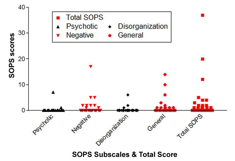
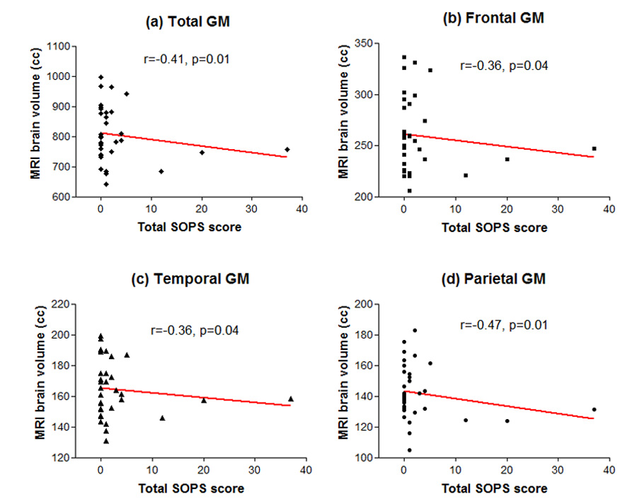
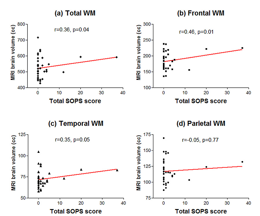
Schizophrenia is characterized by subtle but well-replicated total and regional (frontal and temporal) brain tissue volume deficits. Studies of individuals at-risk for developing schizophrenia suggest that the onset of brain volume decrement may closely pre-date overt manifestations of schizophrenia, making brain volume abnormalities potential predictors for early identification. In an ongoing longitudinal morphometric MRI study of young, nonpsychotic first- or second-degree relatives of schizophrenia probands, we compared brain volumes in 46 relatives who are still within age range for developing schizophrenia against comparison groups of 46 schizophrenia patients and 46 healthy volunteers without family history of schizophrenia. Relatives had similar brain volume abnormalities as schizophrenia patients albeit less severe. Relatives had significantly larger whole brain, frontal, temporal and parietal gray matter (GM) volumes than patients. Relatives also had significantly smaller frontal GM volumes than healthy volunteers. Both relatives and patients had significantly larger whole brain WM (specifically parietal WM) volumes compared to healthy volunteers. Abnormally greater WM volumes in relatives and patients are suggestive of genetically-mediated dysmaturation of the age-expected myelination during adolescence through mid adulthood. On prodromal symptoms assessed in relatives one year after MRI brain scans, initial GM deficits as well as larger WM volumes correlated significantly with greater severity of subsequent prodromal symptoms. Together with previous genetic high-risk studies of adolescent or young adult relatives, these findings indicate that premorbid MRI brain abnormalities may be of predictive value for the early identification of schizophrenia.
Figures
References
-
- Andreasen NC, Endicott J, Spitzer RL, Winokur G. The family history method using diagnostic criteria. Reliability and validity. Arch Gen Psychiatry. 1977;34(10):1229–1235. - PubMed
-
- Andreasen NC, Flaum M, Arndt S. The comprehensive assessment of symptoms and history (cash). An instrument for assessing diagnosis and psychopathology. Arch Gen Psychiatry. 1992;49(8):615–623. - PubMed
-
- Andreasen NC, Rajarethinam R, Cizadlo T, Arndt S, Swayze VW, 2nd, Flashman LA, O'Leary DS, Ehrhardt JC, Yuh WT. Automatic atlas-based volume estimation of human brain regions from mr images. J Comput Assist Tomogr. 1996;20(1):98–106. - PubMed
-
- Baare WF, van Oel CJ, Hulshoff Pol HE, Schnack HG, Durston S, Sitskoorn MM, Kahn RS. Volumes of brain structures in twins discordant for schizophrenia. Arch Gen Psychiatry. 2001;58(1):33–40. - PubMed
-
- Bartzokis G, Nuechterlein KH, Lu PH, Gitlin M, Rogers S, Mintz J. Dysregulated brain development in adult men with schizophrenia: A magnetic resonance imaging study. Biol Psychiatry. 2003;53(5):412–421. - PubMed
Publication types
MeSH terms
Grants and funding
LinkOut - more resources
Full Text Sources
Medical
