. 2007 Nov;18(11):4405-19.
doi: 10.1091/mbc.e07-05-0484. Epub 2007 Aug 29.

Stress-activated protein kinase-mediated down-regulation of the cell integrity pathway mitogen-activated protein kinase Pmk1p by protein phosphatases

Affiliations

Stress-activated protein kinase-mediated down-regulation of the cell integrity pathway mitogen-activated protein kinase Pmk1p by protein phosphatases

Marisa Madrid et al. Mol Biol Cell. 2007 Nov.

Abstract

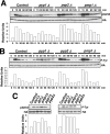
Fission yeast mitogen-activated protein kinase (MAPK) Pmk1p is involved in morphogenesis, cytokinesis, and ion homeostasis as part of the cell integrity pathway, and it becomes activated under multiple stresses, including hyper- or hypotonic conditions, glucose deprivation, cell wall-damaging compounds, and oxidative stress. The only protein phosphatase known to dephosphorylate and inactivate Pmk1p is Pmp1p. We show here that the stress-activated protein kinase (SAPK) pathway and its main effector, Sty1p MAPK, are essential for proper deactivation of Pmk1p under hypertonic stress in a process regulated by Atf1p transcription factor. We demonstrate that tyrosine phosphatases Pyp1p and Pyp2p, and serine/threonine phosphatase Ptc1p, that negatively regulate Sty1p activity and whose expression is dependent on Sty1p-Atf1p function, are involved in Pmk1p dephosphorylation under osmostress. Pyp1p and Ptc1p, in addition to Pmp1p, also control the basal level of MAPK Pmk1p activity in growing cells and associate with, and dephosphorylate Pmk1p both in vitro and in vivo. Our results with Ptc1p provide the first biochemical evidence for a PP2C-type phosphatase acting on more than one MAPK in yeast cells. Importantly, the SAPK-dependent down-regulation of Pmk1p through Pyp1p, Pyp2p, and Ptc1p was not complete, and Pyp1p and Ptc1p phosphatases are able to negatively regulate MAPK Pmk1p activity by an alternative regulatory mechanism. Our data also indicate that Pmk1p phosphorylation oscillates as a function of the cell cycle, peaking at cell separation during cytokinesis, and that Pmp1p phosphatase plays a main role in regulating this process.


Figures

Figure 1.
Figure 1.
The SAPK pathway controls Pmk1p deactivation in growing cells and under hypertonic stress. (A) Schematic arrangement of the main functional components of the SAPK pathway in the fission yeast. RR, response regulator; TF, transcription factor; →, signal transmission; ■ ■ ■, enhanced transcription; ⊥, phosphatase inhibitory effect. See Introduction for details. (B) Strains MI200 (pmk1-HA6H, Control), MI208 (pmk1-HA6H, mcs4Δ), MI 217 (pmk1-HA6H, wak1Δ), MI210 (pmk1-HA6H, wis1Δ), MI204 (pmk1-HA6H, sty1Δ), and MI211 (pmk1-HA6H, atf1Δ) were grown in YES medium to mid-log phase and treated with 0.6 M KCl. Aliquots were harvested at the times indicated, and Pmk1-HA6H was purified by affinity chromatography. Activated Pmk1p was detected by immunoblotting with anti-phospho-p42/44 and total Pmk1p with anti-HA antibodies.
Figure 2.
Figure 2.
Tyrosine phosphatases Pyp1p and Pyp2p are negative regulators of Pmk1p activation during growth and under hypertonic stress. (A) Strains MI200 (pmk1-HA6H, Control), MI213 (pmk1-HA6H, pyp1Δ), MI214 (pmk1-HA6H, pyp2Δ), and MI212 (pmk1-HA6H, pmp1Δ) were grown in YES medium to mid-log phase and treated with 0.6 M KCl. At timed intervals, Pmk1-HA6H was purified by affinity chromatography under native conditions. Activated and total Pmk1p was detected by immunoblotting with anti-phospho-p42/44 or anti-HA antibodies, respectively. (B) The same experiment described in A, except that Pmk1p phosphorylation was detected by immunoblotting with anti-phosphotyrosine (PY99) antibody. (C) Strains MI200 (pmk1-HA6H, Control), MI204 (pmk1-HA6H, sty1Δ), MI211 (pmk1-HA6H, aft1Δ), MI212 (pmk1-HA6H, pmp1Δ), MI213 (pmk1-HA6H, pyp1Δ), and MI214 (pmk1-HA6H, pyp2Δ) were grown in YES medium to mid-log phase, and Pmk1p basal activity was determined with anti-phospho-p42/44 or anti-phosphotyrosine antibodies.
Figure 3.
Figure 3.
Pyp1p and Pyp2p associate with, and dephosphorylate Pmk1p. (A) Strains MI500 (sty1-HA6H, nmt1:GST-pyp1, lanes 1 and 2), MI501 (sty1-HA6H, nmt1:GST-pyp2, lanes 3 and 4), MI502 (pmk1-HA6H, nmt1:GST-pyp1, lanes 5 and 6), MI503 (pmk1-HA6H, nmt1:GST-pyp2, lanes 7 and 8), and MI200 expressing unfused GST from plasmid pDS472a (lanes 9 and 10) were grown in EMM2 medium in the presence (+T) or absence (−T) of thiamine for 16 h. GST, GST-Pyp1, and GST-Pyp2 fusions were purified with glutathione-Sepharose beads, resolved by SDS-PAGE, transferred to nitrocellulose membranes, and immunoblotted with anti-HA or anti-GST antibodies. (B) Pyp1p and Pmk1p associate in vivo. An MI200 transformant expressing unfused GST from plasmid pDS472a (lane 1), and strain MI502 (pmk1-HA6H, pyp1-GST) expressing a genomic version of Pyp1p fused to GST at its C terminus (lane 2) were grown in EMM2 medium in the absence of thiamine for 16 h. GST and Pyp1-GST fusions were purified with glutathione-Sepharose beads, resolved by SDS-PAGE, transferred to nitrocellulose membranes, and immunoblotted with anti-HA or anti-GST antibodies. (C) Pyp1p and Pyp2p dephosphorylate Pmk1p in vitro. Activated Pmk1-HA6H was purified with Ni2+-NTA-agarose beads from strain MI200 after treatment with 0.6 M KCl for 15 min. The beads were incubated at 30°C for 60 min in phosphatase buffer with 5 μg of either GST (lane 1), GST-Pyp2 (lanes 2 and 3), GST-Pyp1 (lanes 4 and 5), or GST-Pmp1 (lanes 6 and 7) in the presence (even lanes) or absence (odd lanes) of 30 mM sodium orthovanadate. The samples were analyzed by SDS-PAGE and immunoblotted with anti-p42/44, anti-HA, and anti-GST antibodies.
Figure 4.
Figure 4.
Ptc1p associates with, and dephosphorylates, Pmk1p. (A) Strains MI200 (pmk1-HA6H, Control), MI216 (pmk1-HA6H, ptc1Δ), MI218 (pmk1-HA6H, ptc2Δ), and MI219 (pmk1-HA6H, ptc3Δ) were grown in YES medium to mid-log phase and treated with 0.6 M KCl. At different times Pmk1-HA6H was purified by affinity chromatography, and either activated or total Pmk1p was detected by immunoblotting with anti-phospho-p42/44 or anti-HA antibodies, respectively. (B) Strains MI200 (pmk1-HA6H, Control), MI212 (pmk1-HA6H, pmp1Δ), MI216 (pmk1-HA6H, ptc1Δ), MI218 (pmk1-HA6H, ptc2Δ), and MI219 (pmk1-HA6H, ptc3Δ) were grown in YES medium to mid-log phase, and Pmk1p basal activity was determined by using anti-phospho-p42/44 antibody as described above. (C) Pmk1p dephosphorylation in vitro. Activated Pmk1-HA6H or Sty1-HA6H was purified with Ni2+-NTA-agarose beads from strains MI200 (Pmk1p; lanes 1–3) or JM1521 (Sty1p; lanes 4–6) after treatment with 0.6 M KCl for 15 min. The beads were incubated at 30°C for 45 min in phosphatase buffer with 10 μg of GST-Ptc1 (lanes 2, 3, 5, and 6) in the presence of 10 mM MgCl2 (lanes 3 and 5) or 10 mM EDTA (lanes 2 and 6). The samples were analyzed by SDS-PAGE and immunoblotted with anti-p42/44, anti-HA, and anti-GST antibodies. (D) Pmk1p and Ptc1p interact in vitro. Cell lysates from exponentially growing strains JM1521 (Sty1-HA6H; lanes 1 and 3), MI200 (Pmk1-HA6H; lanes 2 and 4), and strain MI200 subjected to a 15-min treatment with 0.6 M KCl (lane 5) were incubated each with 15 μg of bacterially purified GST (lanes 1 and 2) or GST-Ptc1 (lanes 3–5) bound to glutathione-Sepharose beads. The beads were washed extensively, and the binding of Pmk1p or Sty1p fusions was detected by inmunoblotting with monoclonal anti-HA antibodies. (E) Pmk1p and Ptc1p interact in vivo. An MI200 transformant expressing unfused GST from plasmid pDS472a (lane 1) and strain MI505 (pmk1-HA6H, ptc1-GST) expressing a genomic version of Ptc1p fused to GST at its C terminus (lane 2) were grown in EMM2 medium in the absence of thiamine for 16 h. GST and Pyp1-GST fusions were purified with glutathione-Sepharose beads, resolved by SDS-PAGE, transferred to nitrocellulose membranes, and immunoblotted with anti-HA or anti-GST antibodies.
Figure 5.
Figure 5.
Sty1p hyperactivation promotes defective Pmk1p activation under saline stress. (A) Sty1p basal activity in different mutants. Strains JM1521 (sty1-HA6H, Control), MI1001 (sty1-HA6H, pyp1Δ), MI1002 (sty1-HA6H, pyp2Δ), MI1003 (sty1-HA6H, ptc1Δ), MI1000 (sty1-HA6H, pmp1Δ), and MI100 (sty1-HA6H, pmk1Δ) were grown in YES medium to mid-log phase, and Sty1-HA6H was purified by affinity chromatography. Activated or total Sty1p was detected by immunoblotting with anti-phospho-p38 and anti-HA antibodies, respectively. (B) Increased Pyp2p and Ptc1p synthesis in pyp1Δ cells. Strains MI702 (pyp2-13myc, Control) and MI704 (pyp2-13myc, pyp1Δ) (top), MI703 (ptc1-13myc, Control), and MI705 (ptc1-13myc, pyp1Δ) (bottom) were grown in YES medium to mid-log phase and treated with 0.6 M KCl. Aliquots were harvested at the times indicated, and total extracts were obtained. Pyp2-13myc and Ptc1p-myc fusions were detected by immunoblotting with anti-c-myc antibody. Anti-Cdc2 antibody was used as loading control. (C) Pmk1p activation in wis1DD cells. Strains MI200 (pmk1-HA6H, Control), MI709 (pmk1-HA6H, wis1DD), and MI713 (pmk1-HA6H, wis1DD, atf1Δ) were grown in YES medium to mid-log phase and treated with 0.6 M KCl. Aliquots were harvested at the times indicated and Pmk1-HA6H was purified by affinity chromatography. Activated Pmk1p was detected by immunoblotting with anti-phospho-p42/44 and total Pmk1p with anti-HA antibodies. (D) Increased Pyp1p, Pyp2p and Ptc1p synthesis in wis1DD cells. Strains MI701 (pyp1-13myc, Control) and MI710 (pyp1-13myc, wis1DD) (top), MI702 (pyp2-13myc, Control) and MI711 (pyp2-13myc, wis1DD) (middle), MI703 (ptc1-13myc, Control) and MI712 (ptc1-13myc, wis1DD) (bottom) were grown in YES medium to mid-log phase and treated with 0.6 M KCl. Aliquots were harvested at the times indicated, and total extracts were prepared. Pyp1-myc, Pyp2-13myc, and Ptc1p-myc fusions were detected by immunoblotting with anti-c-myc antibody. Anti-Cdc2 antibody was used as loading control.
Figure 6.
Figure 6.
Synergistic down-regulation of Pmk1p activity by Pyp1p and Ptc1p phosphatases. (A) Pmk1p basal activity in phosphatase-null mutants. Pmk1-HA6H was purified by affinity chromatography from strains MI200 (pmk1-HA6H, Control), MI213 (pmk1-HA6H, pyp1Δ), MI216 (pmk1-HA6H, ptc1Δ), and MI223 (pmk1-HA6H, pyp1Δ, ptc1Δ). Activated or total Pmk1p was detected by immunoblotting with anti-phospho-p42/44 or anti-HA antibodies, respectively. (B) Pmk1p activation in phosphatase-null mutants subjected to a saline stress. Strains MI200 (pmk1-HA6H, Control), MI223 (pmk1-HA6H, pyp1Δ, ptc1Δ), and MI213 (pmk1-HA6H, pyp1Δ) were grown in YES medium to mid-log phase and treated with 0.6 M KCl. At different times Pmk1-HA6H was purified by affinity chromatography, and either activated or total Pmk1p was detected by immunoblotting as described above.
Figure 7.
Figure 7.
Pyp1p and Ptc1p phosphatases can negatively modulate Pmk1p phosphorylation under osmotic stress in a Sty1p-independent manner. (A) Strains MI200 (pmk1-HA6H, Control), MI204 (pmk1-HA6H, sty1Δ), MI220 (pmk1-HA6H, sty1Δ, pyp1Δ), MI221 (pmk1-HA6H, sty1Δ, pyp2Δ), and MI222 (pmk1-HA6H, sty1Δ, ptc1Δ) were grown in YES medium to mid-log phase and treated with 0.6 M KCl. At timed intervals, Pmk1-HA6H was purified by affinity chromatography under native conditions, and activated or total Pmk1p was detected by immunoblotting with anti-phospho-p42/44 or anti-HA antibodies, respectively. (B) Synthesis of Pyp1p and Ptc1p phosphatases in sty1Δ cells. Strains MI701 (pyp1-13myc, Control) and MI706 (pyp1-13myc, sty1Δ) (left), MI702 (pyp2-13myc, Control) and MI707 (pyp2-13myc, sty1Δ) (middle), MI703 (ptc1-13myc, Control) and MI708 (ptc1-13myc, sty1Δ) (right) were grown in YES medium to mid-log phase and treated with 0.6 M KCl. Aliquots were harvested at the times indicated, and total extracts prepared. Pyp1-myc, Pyp2-13myc, and Ptc1p-myc fusions were detected by immunoblotting with anti-c-myc antibody. Anti-Cdc2 antibody was used as loading control.
Figure 8.
Figure 8.
Differential regulation of MAPK functions by Pyp1p, Ptc1p, and Pmp1p. (A) Chloride sensitivity assays. Strains MI200 (Control), TK107 (sty1Δ), MI102 (pmk1Δ), MI107 (pmk1Δ sty1Δ), MI104 (pmp1Δ), MI116 (pmp1Δ sty1Δ), MI115 (pmp1Δ pmk1Δ), MI105 (pyp1Δ), MI109 (pyp1Δ sty1Δ), MI108 (pyp1Δ pmk1Δ), MI106 (pyp2Δ), MI118 (pyp2Δ sty1Δ), MI117 (pyp2Δ pmk1Δ), MI110 (ptc1Δ), MI112 (ptc1Δ sty1Δ), MI111 (ptc1Δ pmk1Δ), and 2119 (wis1DD) were grown in YES medium to an A600 of 0.5. Samples containing 105, 104, 103 or 102 cells were spotted onto YES plates supplemented with 0.2 M MgCl2 and incubated for 3 d at 28°C before being photographed. (B) Cell wall integrity assays. Strains MI200 (Control), TK107 (sty1Δ), MI102 (pmk1Δ), MI104 (pmp1Δ), MI116 (pmp1Δ sty1Δ), MI115 (pmp1Δ pmk1Δ), MI105 (pyp1Δ), MI109 (pyp1Δ sty1Δ), MI108 (pyp1Δ pmk1Δ), MI110 (ptc1Δ), MI112 (ptc1Δ sty1Δ), and MI111 (ptc1Δ pmk1Δ) were grown in YES medium to an A600 of 0.5, and the cells treated at 30°C with 100 μg/ml Zymolyase 20-T. Cell lysis was monitored by measuring A600 decay at different incubation times. Results represent the mean value of three independent experiments.
Figure 9.
Figure 9.
Influence of Pmp1p activity on the cell cycle-dependent activation of Pmk1p. (A) Pmk1p activates periodically during the cell cycle. Top, cells from strains MI600 (cdc25-22, pmk1-HA6H) and MI602 (cdc25-22, pmp1Δ, pmk1-HA6H) were grown to an A600 of 0.3 at 25°C, shifted to 37°C for 3.5 h, and then released from the growth arrest by transfer back to 25°C. Aliquots were taken at different time intervals, and Pmk1-HA6H was purified by affinity chromatography. Activated or total Pmk1p were detected by immunoblotting with anti-phospho-p42/44 or anti-HA antibodies, respectively, and anti-Cdc2 antibody was used as loading control. Bottom, septation index of strains MI600 (filled circles), MI601 (cdc25-22, pmk1Δ; open circles), and MI602 (filled triangles). (B) Quantitative analysis of Pmk1p activity during cell cycle in strains MI600 (filled bars) and MI602 (empty bars) obtained from data in A. (C) Defective cell separation in pmk1Δ and pmp1Δ cells. Cells from strains MI600 (Control), MI601, and MI602 were taken at the times shown during cell cycle experiments, stained with Calcofluor white, and observed by fluorescence microscopy. Arrows indicate cells with multiseptate phenotype and defective cell separation.
Figure 10.
Figure 10.
Proposed regulatory links between the SAPK and the cell integrity pathways by protein phosphatases (shaded figures) during growth and osmostress. Pyp1p, Pyp2p, and Ptc1p protein phosphatases are negative regulators of MAPK Sty1p and MAPK Pmk1 activities, whereas Pmp1 only dephosphorylates MAPK Pmk1p (⊥, inhibition). The expression/synthesis of the shared phosphatases is in turn under Sty1p/Atf1p-dependent and Sty1p/Atf1p-independent (X-dependent) regulation. The size of the discontinuous arrows indicates the relative transcriptional reliance. The question mark shows that the corresponding transcription factor is unknown. For more details on abbreviations and symbols, see Figure 1.

Similar articles

Cited by

References

    1. Alfa C., Fantes P., Hyams J., Mcleod M., Warbrick E. New York: Cold Spring Harbor Laboratory Press; 1993. Experiments with Fission Yeast: A Laboratory Course Manual.
    1. Bahler J., Wu J. Q., Longtine M. S., Shah N. G., McKenzie A., 3rd, Steever A. B., Wach A., Philippsen P., Pringle J. R. Heterologous modules for efficient and versatile PCR-based gene targeting in Schizosaccharomyces pombe. Yeast. 1998;14:943–951. - PubMed
    1. Bone N., Millar J. B., Toda T., Armstrong J. Regulated vacuole fusion and fission in Schizosaccharomyces pombe: an osmotic response dependent on MAP kinases. Curr. Biol. 1998;8:135–144. - PubMed
    1. Chen D., Toone W. M., Mata J., Lyne R., Burns G., Kivinen K., Brazma A., Jones N., Bähler J. Global transcriptional responses of fission yeast to environmental stress. Mol. Biol. Cell. 2003;14:214–229. - PMC - PubMed
    1. Degols G., Shiozaki K., Russell P. Activation and regulation of the Spc1 stress-activated protein kinase in Schizosaccharomyces pombe. Mol. Cell. Biol. 1996;16:2870–2877. - PMC - PubMed

Publication types

MeSH terms

Substances

LinkOut - more resources