. 2007 Nov 15;176(10):974-82.
doi: 10.1164/rccm.200702-334OC. Epub 2007 Aug 29.

Transforming growth factor-beta1 suppresses airway hyperresponsiveness in allergic airway disease

Affiliations

Transforming growth factor-beta1 suppresses airway hyperresponsiveness in allergic airway disease

John F Alcorn et al. Am J Respir Crit Care Med. .

Abstract

Rationale: Asthma is characterized by increases in airway resistance, pulmonary remodeling, and lung inflammation. The cytokine transforming growth factor (TGF)-beta has been shown to have a central role in asthma pathogenesis and in mouse models of allergic airway disease.

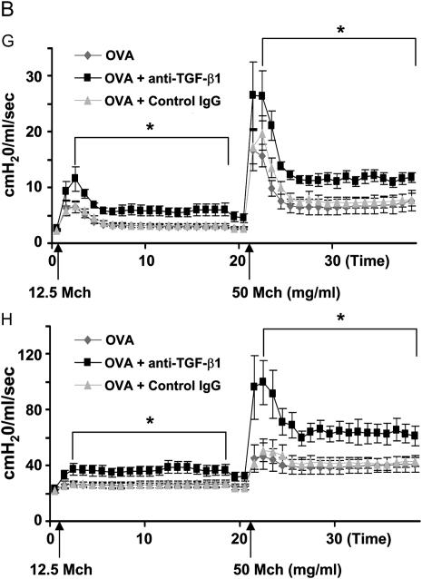
Objectives: To determine the contribution of TGF-beta to airway hyperresponsiveness (AHR), we examined the time course, source, and isoform specificity of TGF-beta production in an in vivo mouse asthma model. To then elucidate the function of TGF-beta in AHR, inflammation, and pulmonary fibrosis, we examined the effects of blocking TGF-beta signaling with neutralizing antibody.

Methods: Mice were sensitized and challenged with ovalbumin (OVA) to establish allergic airway disease. TGF-beta activity was neutralized by intranasal administration of monoclonal antibody.

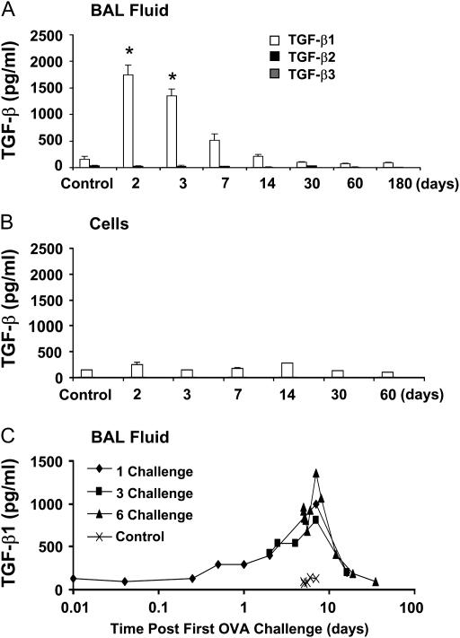
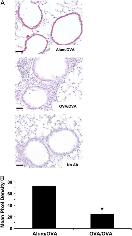
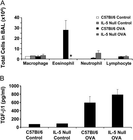
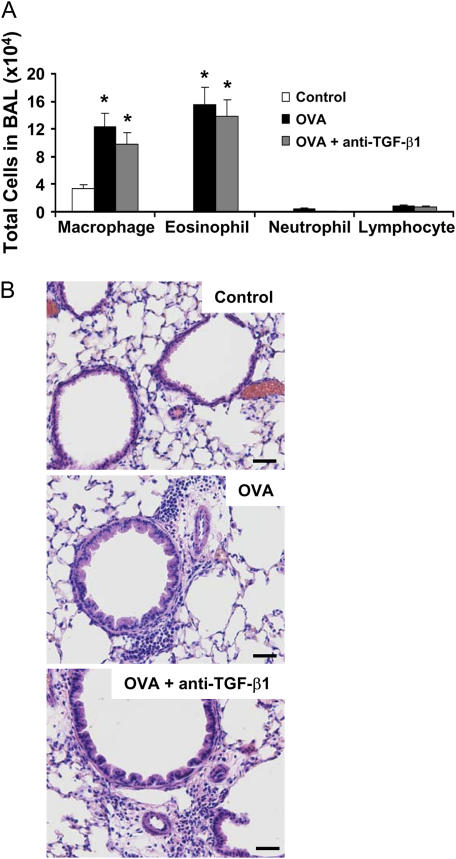
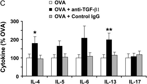
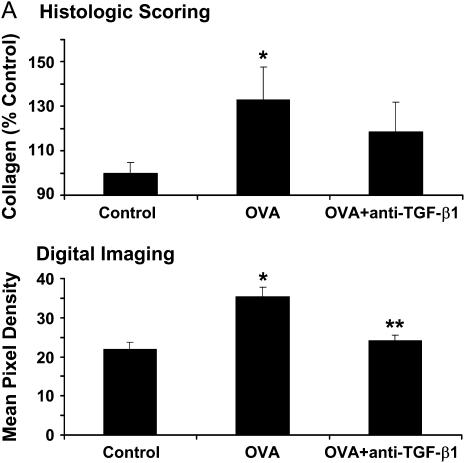
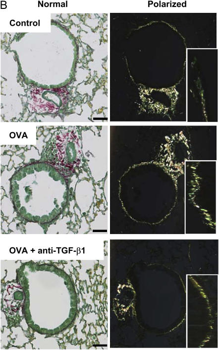
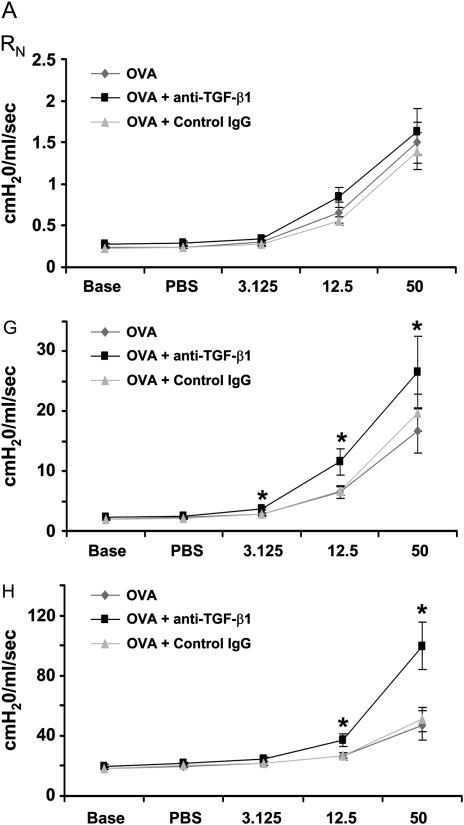
Measurements and main results: TGF-beta1 protein levels were increased in OVA-challenged lungs versus naive controls, and airway epithelial cells were shown to be a likely source of TGF-beta1. In addition, TGF-beta1 levels were elevated in OVA-exposed IL-5-null mice, which fail to recruit eosinophils into the airways. Neutralization of TGF-beta1 with specific antibody had no significant effect on airway inflammation and eosinophilia, although anti-TGF-beta1 antibody enhanced OVA-induced AHR and suppressed pulmonary fibrosis.

Conclusions: These data show that TGF-beta1 is the main TGF-beta isoform produced after OVA challenge, with a likely cellular source being the airway epithelium. The effects of blocking TGF-beta1 signaling had differential effects on AHR, fibrosis, and inflammation. While TGF-beta neutralization may be beneficial to abrogating airway remodeling, it may be detrimental to lung function by increasing AHR.


Figures

<b>Figure 1.</b>
Figure 1.
Detection of transforming growth factor (TGF)-β isoforms in the bronchoalveolar lavage (BAL) fluid after ovalbumin (OVA) sensitization and challenge. BALB/c mice were sensitized to OVA and challenged with 1% aerosolized OVA on 6 consecutive days, and BAL samples were analyzed at the indicated time points after the last challenge (n = 4 mice each). Control mice were not sensitized or challenged with OVA. TGF-β levels were measured by isoform-specific ELISA on the BAL supernatant (A) or cell lysate (B). TGF-β1 levels in the BAL fluid were measured at the indicated times after one, three, or six OVA challenges (C). *P < 0.05.
<b>Figure 2.</b>
Figure 2.
Transforming growth factor (TGF)-β1 protein colocalizes with the bronchial epithelium and is decreased upon ovalbumin (OVA) sensitization and challenge. BALB/c mice were challenged six times with 1% aerosolized OVA, and paraffin-embedded sections were prepared. Immunohistochemistry specific for TGF-β1 was then performed as described in Methods (A) (n = 5 mice each). Alum/OVA indicates control mice that were sham sensitized then challenged with OVA, OVA/OVA indicates OVA-sensitized and -challenged mice, No Ab indicates a no primary antibody control. TGF-β1 is indicated by red staining at ×200 original magnification; scale bar equals 25 μm. TGF-β1 staining was then quantified by mean pixel density (B) (n = 12), OVA/OVA-treated mice had decreased TGF-β1 immunoreactivity compared with Alum/OVA. *P < 0.05.
<b>Figure 3.</b>
Figure 3.
IL-5–null mice produce similar transforming growth factor (TGF)-β1 levels to control animals after ovalbumin (OVA) challenge despite decreased eosinophilia. IL-5–null mice or C57BL/6 controls were sensitized to OVA and challenged six times with 1% aerosolized OVA. Bronchoalveolar lavage (BAL) samples were then taken and analyzed for cell number, identity, and TGF-β1 levels (n = 3 mice each). IL-5–null mice failed to recruit eosinophils into the airspaces compared with controls (A). Both control and IL-5–null mice produce similar levels of TGF-β1 protein in the BAL fluid (B). *P = 0.058.
<b>Figure 4.</b>
Figure 4.
Ovalbumin (OVA)-induced phosphorylation of Smad 2 is attenuated by anti–transforming growth factor (TGF)-β1 antibody treatment. BALB/c mice were sensitized to OVA and challenged on 3 consecutive days with 1% aerosolized OVA. TGF-β1 neutralizing antibodies (75 μg) or control nonspecific IgG1 were administered once daily for 4 days starting the day of the first OVA challenge. Paraffin-embedded lung sections were then stained for phospho-Smad 2 (red) and counterstained with the nuclear dye Sytox Green (green) (A). Nuclear phospho-Smad 2 appears yellow due to the overlapping stains. Images were taken at ×200 original magnification by confocal microscopy; scale bar equals 25 μm. Phospho-Smad 2 staining was then quantified by mean pixel density (B) (n = 21, 24, 24, respectively). Anti–TGF-β1 significantly decreased phospho-Smad 2 immunoreactivity. *P < 0.05.
<b>Figure 5.</b>
Figure 5.
Neutralization of transforming growth factor (TGF)-β1 does not alter pulmonary inflammation in mice subjected to ovalbumin (OVA) sensitization and challenge. BALB/c mice were sensitized to OVA and challenged on 3 consecutive days with 1% aerosolized OVA. TGF-β1 neutralizing antibodies (75 μg) were administered once daily for 4 days starting the day of the first OVA challenge. Cell number and identity were determined in the bronchoalveolar lavage (BAL) (n = 4, 8, 9 mice, respectively) (A). Whole lung sections were prepared by paraffin embedding and stained with hematoxylin and eosin to examine the tissue inflammation in the treatment groups (B), ×200 original magnification. BAL cytokine profiles were assessed by Bioplex analysis (n = 7, 8, 8, respectively) (C). *P < 0.06 versus OVA mice; **P < 0.05 versus OVA mice.
<b>Figure 6.</b>
Figure 6.
Neutralization of transforming growth factor (TGF)-β1 decreases subepithelial collagen deposition after ovalbumin (OVA) challenge. BALB/c mice were sensitized to OVA and challenged on 3 consecutive days with 1% aerosolized OVA. TGF-β1 neutralizing antibodies (75 μg) were administered once daily for 4 days starting the day of the first OVA challenge. Picosirius red–stained paraffin sections were assessed by blinded scorers for collagen deposition (A, top) (n = 6, 12, 15 mice, respectively). Picosirius red images were then analyzed by mean pixel density for collagen deposition (A, bottom) (n = 27, 27, 30 airways analyzed, respectively). Differential interference contrast images of Picosirius red–stained lung sections (B, ×200 original magnification, scale bar = 25 μm). Insets depict a portion of the airway wall at increased magnification. (A, top) *P = 0.059 versus control mice (histologic scoring); (A, bottom) *P < 0.05 versus control; **P < 0.05 versus OVA (digital imaging); OVA + anti–TGF-β1 were not statistically different from control.
<b>Figure 7.</b>
Figure 7.
Neutralization of transforming growth factor (TGF)-β1 enhances ovalbumin (OVA)-induced airway hyperresponsiveness (AHR). BALB/c mice were sensitized to OVA and challenged on 3 consecutive days with 1% aerosolized OVA. TGF-β1 neutralizing antibodies (75 μg) were administered once daily for 4 days starting the day of the first OVA challenge. After challenge, airway function was assessed by Flexivent (SCIREQ, Montreal, PQ, Canada). TGF-β1 neutralization enhanced OVA sensitization and challenge induced AHR (A) (peak response; n = 7, 8, 8 mice, respectively). In addition to increasing peak methacholine (Mch) responses, TGF-β1 antibody enhanced prolonged AHR between Mch doses (B). There were no effects of control IgG treatment on OVA-induced AHR. AHR as measured by the G (tissue resistance) and H (tissue elastance) parameters were significantly increased. *P < 0.05 versus OVA mice.

Similar articles

Cited by

References

    1. Leask A, Abraham DJ. TGF-beta signaling and the fibrotic response. FASEB J 2004;18:816–827. - PubMed
    1. Bartram U, Speer CP. The role of transforming growth factor beta in lung development and disease. Chest 2004;125:754–765. - PubMed
    1. Kumar RK, Herbert C, Foster PS. Expression of growth factors by airway epithelial cells in a model of chronic asthma: regulation and relationship to subepithelial fibrosis. Clin Exp Allergy 2004;34:567–575. - PubMed
    1. Minshall EM, Leung DY, Martin RJ, Song YL, Cameron L, Ernst P, Hamid Q. Eosinophil-associated TGF-β1 mRNA expression and airways fibrosis in bronchial asthma. Am J Respir Cell Mol Biol 1997;17:326–333. - PubMed
    1. Tanaka H, Komai M, Nagao K, Ishizaki M, Kajiwara D, Takatsu K, Delespesse G, Nagai H. Role of interleukin-5 and eosinophils in allergen-induced airway remodeling in mice. Am J Respir Cell Mol Biol 2004;31:62–68. - PubMed

Publication types

MeSH terms