. 2007 Nov;27(11):2484-90.
doi: 10.1161/ATVBAHA.107.151100. Epub 2007 Aug 30.

Segregation of platelet aggregatory and procoagulant microdomains in thrombus formation: regulation by transient integrin activation

Affiliations

Segregation of platelet aggregatory and procoagulant microdomains in thrombus formation: regulation by transient integrin activation

Imke C A Munnix et al. Arterioscler Thromb Vasc Biol. 2007 Nov.

Abstract

Objective: Platelets play a dual role in thrombosis by forming aggregates and stimulating coagulation. We investigated the commitment of platelets to these separate functions during collagen-induced thrombus formation in vitro and in vivo.

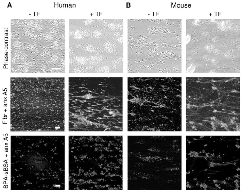
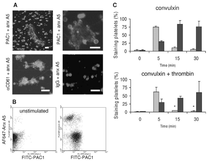
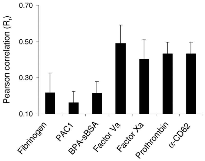
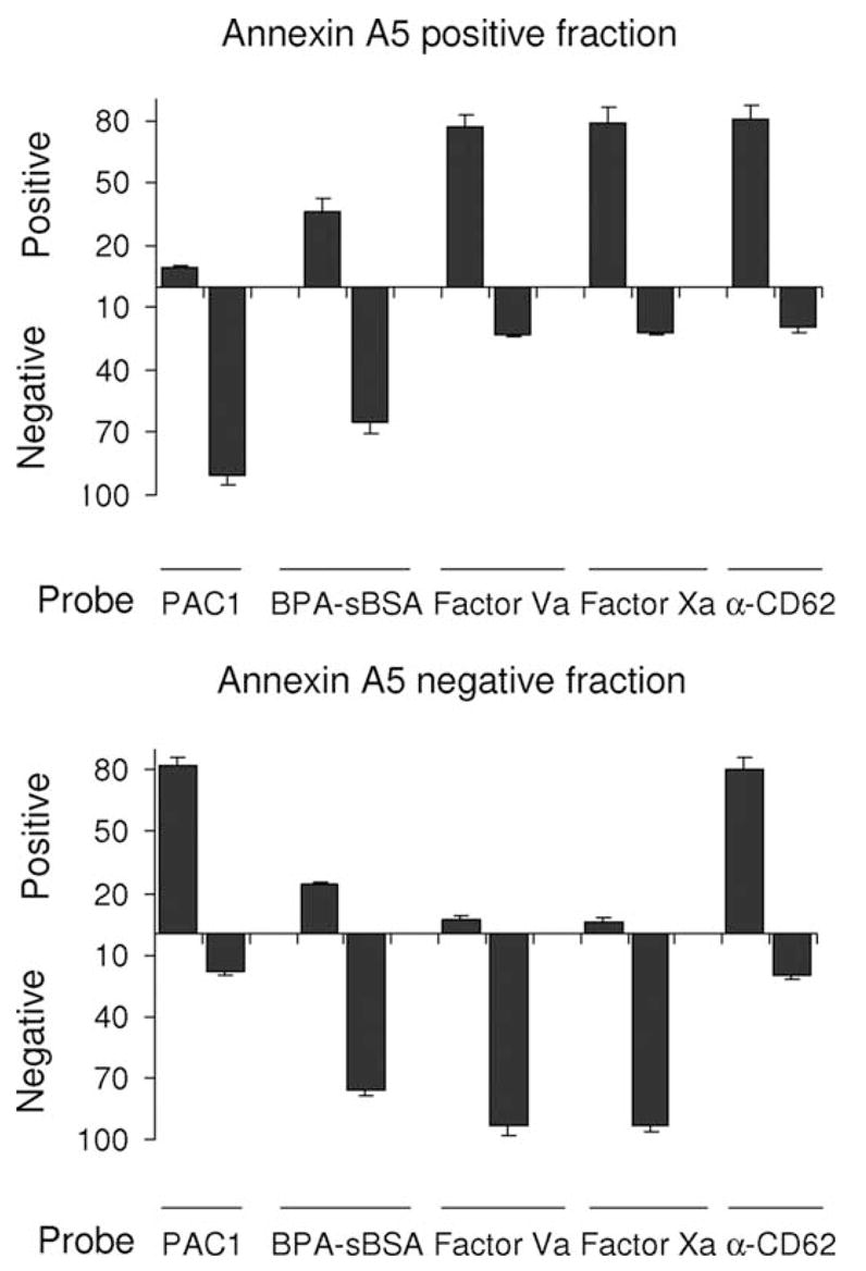
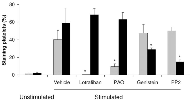
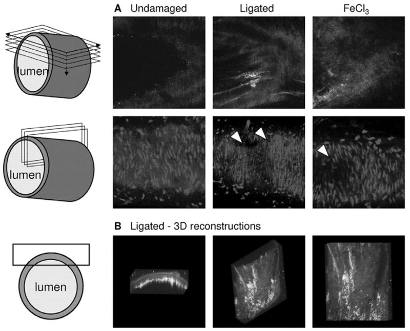
Methods and results: High-resolution 2-photon fluorescence microscopy revealed that in thrombus formation under flow, fibrin(ogen)-binding platelets assembled into separate aggregates, whereas distinct patches of nonaggregated platelets exposed phosphatidylserine. The latter platelet population had inactivated alphaIIb beta3 integrins and displayed increased binding of coagulation factors. Coated platelets, expressing serotonin binding sites, were not identified as a separate population. Thrombin generation and coagulation favored the transformation to phosphatidylserine-exposing platelets with inactivated integrins and reduced adhesion. Prolonged tyrosine phosphorylation in vitro resulted in secondary downregulation of active alphaIIb beta3.

Conclusions: These results lead to a new spatial model of thrombus formation, in which aggregated platelets ensure thrombus stability, whereas distinct patches of nonaggregated platelets effectuate procoagulant activity and generate thrombin and fibrin. Herein, the hemostatic activity of a developing thrombus is determined by the balance in formation of proaggregatory and procoagulant platelets. This balance is influenced by antiplatelet and anticoagulant medication.


Figures

Figure 1
Figure 1
Heterogeneity in human and murine thrombi formed on collagen. Flow experiments were performed with human (A) or murine (B) blood in the absence of tissue factor (−TF) using PPACK-anticoagulated blood (left columns); or with citrate blood that was perfused together with tissue factor (+TF, 2 pM, f.c.) and CaCl2 (2 mmol/L free Ca2+, f.c.) to allow coagulation (right columns). Standard perfusion time was 4 minutes at a shear rate of 1000 s −1. Blood was preincubated with 0.2 mg/mL OG-fibrinogen. Alternatively, preincubation was with 50 μg/mL BPA-sBSA and postlabeling with 1 μg/mL AF532-labeled streptavidin. In both cases, AF647-annexin A5 was also present. Upper panels, Bright-field phase-contrast images after perfusion. Middle panels, TPLSM images of OG-fibrinogen (green) and AF647-annexin A5 (red) fluorescence (different fields of view). Lower panels, TPLSM images of BPA-sBSA (blue) and AF647-annexin A5 (red) staining. Images are representative of 4 to 8 experiments; bars indicate 20 μm.
Figure 2
Figure 2
Temporary integrin activation state in procoagulant platelets. A, Human thrombi were formed on collagen in the presence of tissue factor/CaCl2 (see Figure 1). Blood was labeled with 0.5 μg/mL AF647-annexin A5 plus either 0.6 μg/mL FITC-PAC1, 1.25 μg/mL FITC-α-CD61, or 1.25 μg/mL FITC-IgG. Representative TPLSM images are shown of FITC (green) and AF647 (red) fluorescence at lower and higher magnifications. Optical zoom 1 to 4×; bars indicate 20 μm. B and C, Washed platelets were stimulated in the presence of 2 mmol/L CaCl2 with 50 ng/mL convulxin alone or with 4 nmol/L thrombin, for 5 to 30 minutes without stirring (1 × 10⁷ platelets/mL). B, Representative dot plots are given of FL1 (FITC) vs FL4 (AF647) after 5 minutes of stimulation with convulxin. C, Two-color flow cytometry after costaining at indicated times with FITC-PAC1 and AF647-annexin A5, showing transient appearance of PAC1 binding sites and persistent PS exposure. Data are percentages of platelets staining with FITC-PAC1 (gray bars) or OG-annexin A5 (black bars). Platelets were selected according to their forward/side scatter characteristics. Mean ± SD (n = 4), *P < 0.05 compared with t = 5 minutes.
Figure 3
Figure 3
Partial overlap of binding of coagulation factors and annexin A5 to platelets in thrombi. Human thrombi were generated on collagen under flow and coagulant conditions (see Figure 1) in the presence of AF647-annexin A5 (red) plus either 20 nmol/L AF488-factor Va, 200 nmol/L OG-prothrombin, 100 nmol/L OG-factor Xa, or 1.25 μg/mL FITC-α-CD62 mAb (green). Histograms give pattern overlap analysis of TPLSM images for annexin A5 (red) and indicated factor or antibody (green); shown is the Pearson correlation coefficient (Rr) of corresponding green and red bit maps (n = 4 to 7).
Figure 4
Figure 4
Heterogeneity in binding properties of PS-exposing platelets. Platelets in suspension were either unstimulated or stimulated with 50 ng/mL convulxin, 4 nmol/L thrombin and 2 mmol/L CaCl2. Two-color flow cytometry was performed in the presence of AF647-annexin A5 combined with either 20 nmol/L AF488-factor Va, 100 nmol/L OG-factor Xa, 200 nmol/L OG-prothrombin, or 1.25 μg/mL FITC- α-CD62 mAb. Histograms show binding properties of the annexin A5 positive and negative platelet fractions after 10 minutes of activation: percentages of cells staining positively (upside bars) or negatively (downside bars) with indicated green probe: FITC-PAC1, AF532-streptavidin BPA-sBSA, AF488-factor Va, OG-factor Xa, or FITC-α-CD62. Mean ± SD (n = 4).
Figure 5
Figure 5
Modulation of protein tyrosine phosphorylation alters integrin activation and PS exposure. Human washed platelets were stimulated with 50 ng/mL convulxin and 4 nmol/L thrombin in the presence of 2 mmol/L CaCl2. The cells were preincubated for 10 minutes with vehicle, 200 μmol/L phenylarsine oxide (PAO), 100 μmol/L genistein, 20 μmol/L PP2, or 10 μmol/L lotrafiban. Colabeling was with a mixture of FITC-PAC1 and AF647-annexin A5. Data are expressed as percentages of platelets staining with FITC-PAC1 (gray bars) or OG-annexin A5 (black bars). Mean ± SD (n = 4), *P < 0.05 compared with stimulation in the presence of vehicle.
Figure 6
Figure 6
Heterogeneity in arterial thrombi induced by carotid ligation or by FeCl3 application. Mice were infused with OG-fibrinogen and AF568-annexin A5 (200 μg each). A, left panels, Undamaged control carotid artery. Middle panels, One carotid artery was damaged by tight ligation at the bifurcation for 5 minutes to induce vascular damage. Right panels, In other animals, 1 carotid artery was damaged by local application of saturated FeCl3. Thrombus formation proceeded for 10 minutes, after which fluorescence at the luminal side of the artery was recorded by TPLSM. Upper row, Images of fibrin(ogen) (green) and annexin A5 (red) fluorescence at the damaged side of the vessel wall. Note distinct patches of green and red fluorescence; also note blue autofluorescence, indicating the demarcation of the vessel wall with the lumen. Second row, Images of Syto-44 fluorescence (blue) at cross section through the vessel wall. Arrow heads indicate absence of cell nuclei at side of damage. B, Three-dimensional reconstruction of fibrin(ogen) and annexin A5 fluorescence within the ligated artery: side view through vessel wall, turned view, and view from inside of vessel lumen. Data are representative of 3 to 4 vessels. Cartoons indicate way of optical sectioning (images 206 × 206 μm).

References

    1. Heemskerk JWM, Kuijpers MJE, Munnix ICA, Siljander PRM. Platelet collagen receptors and coagulation. Trends Cardiovasc Med. 2005;15:86–92. - PubMed
    1. Zwaal RFA, Schroit AJ. Pathophysiologic implications of membrane phospholipid assymetry in blood cells. Blood. 1997;89:1121–1132. - PubMed
    1. Munnix ICA, Harmsma M, Giddings JC, Collins PW, Feijge MAH, Comfurius P, Heemskerk JWM, Bevers EM. Store-mediated calcium entry in the regulation of phosphatidylserine exposure in blood cells from Scott patients. Thromb Hemost. 2003;89:687–695. - PubMed
    1. Butenas S, van ’t Veer C, Mann KG. ‘Normal’ thrombin generation. Blood. 1999;94:2169–2178. - PubMed
    1. Monroe DM, Hoffman M, Roberts HR. Platelets and thrombin generation. Arterioscler Thromb Vasc Biol. 2002;22:1381–1389. - PubMed

Publication types

Substances