Fibrin gel-immobilized VEGF and bFGF efficiently stimulate angiogenesis in the AV loop model
- PMID: 17762899
- PMCID: PMC1960744
- DOI: 10.2119/2007-00057.Arkudas
Fibrin gel-immobilized VEGF and bFGF efficiently stimulate angiogenesis in the AV loop model
Abstract
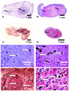
The modulation of angiogenic processes in matrices is of great interest in tissue engineering. We assessed the angiogenic effects of fibrin-immobilized VEGF and bFGF in an arteriovenous loop (AVL) model in 22 AVLs created between the femoral artery and vein in rats. The loops were placed in isolation chambers and were embedded in 500 microL fibrin gel (FG) (group A) or in 500 microL FG loaded with 0.1 ng/microL VEGF and 0.1 ng/microL bFGF (group B). After two and four weeks specimens were explanted and investigated using histological, morphometrical, and ultramorphological [scanning electron microscope (SEM) of vascular corrosion replicas] techniques. In both groups, the AVL induced formation of densely vascularized connective tissue with differentiated and functional vessels inside the fibrin matrix. VEGF and bFGF induced significantly higher absolute and relative vascular density and a faster resorption of the fibrin matrix. SEM analysis in both groups revealed characteristics of an immature vascular bed, with a higher vascular density in group B. VEGF and bFGF efficiently stimulated sprouting of blood vessels in the AVL model. The implantation of vascular carriers into given growth factor-loaded matrix volumes may eventually allow efficient generation of axially vascularized, tissue-engineered composites.
Figures


Similar articles
-
The impact of VEGF and bFGF on vascular stereomorphology in the context of angiogenic neo-arborisation after vascular induction.J Electron Microsc (Tokyo). 2011;60(4):267-74. doi: 10.1093/jmicro/dfr025. Epub 2011 May 28. J Electron Microsc (Tokyo). 2011. PMID: 21622976
-
Dose-finding study of fibrin gel-immobilized vascular endothelial growth factor 165 and basic fibroblast growth factor in the arteriovenous loop rat model.Tissue Eng Part A. 2009 Sep;15(9):2501-11. doi: 10.1089/ten.tea.2008.0477. Tissue Eng Part A. 2009. PMID: 19292678
-
Angiogenic synergistic effect of basic fibroblast growth factor and vascular endothelial growth factor in an in vitro quantitative microcarrier-based three-dimensional fibrin angiogenesis system.World J Gastroenterol. 2004 Sep 1;10(17):2524-8. doi: 10.3748/wjg.v10.i17.2524. World J Gastroenterol. 2004. PMID: 15300897 Free PMC article.
-
Modified fibrin hydrogel matrices: both, 3D-scaffolds and local and controlled release systems to stimulate angiogenesis.Curr Pharm Des. 2007;13(35):3597-607. doi: 10.2174/138161207782794158. Curr Pharm Des. 2007. PMID: 18220797 Review.
-
Flow-Induced Axial Vascularization: The Arteriovenous Loop in Angiogenesis and Tissue Engineering.Plast Reconstr Surg. 2016 Oct;138(4):825-835. doi: 10.1097/PRS.0000000000002554. Plast Reconstr Surg. 2016. PMID: 27673517 Review.
Cited by
-
Flow increase is decisive to initiate angiogenesis in veins exposed to altered hemodynamics.PLoS One. 2015 Jan 30;10(1):e0117407. doi: 10.1371/journal.pone.0117407. eCollection 2015. PLoS One. 2015. PMID: 25635764 Free PMC article.
-
A Low Cost Implantation Model in the Rat That Allows a Spatial Assessment of Angiogenesis.Front Bioeng Biotechnol. 2018 Feb 5;6:3. doi: 10.3389/fbioe.2018.00003. eCollection 2018. Front Bioeng Biotechnol. 2018. PMID: 29468155 Free PMC article.
-
Evaluation of angiogenesis of bioactive glass in the arteriovenous loop model.Tissue Eng Part C Methods. 2013 Jun;19(6):479-86. doi: 10.1089/ten.TEC.2012.0572. Epub 2013 Jan 16. Tissue Eng Part C Methods. 2013. PMID: 23189952 Free PMC article.
-
Hyaluronan-based heparin-incorporated hydrogels for generation of axially vascularized bioartificial bone tissues: in vitro and in vivo evaluation in a PLDLLA-TCP-PCL-composite system.J Mater Sci Mater Med. 2011 May;22(5):1279-91. doi: 10.1007/s10856-011-4300-0. Epub 2011 Mar 30. J Mater Sci Mater Med. 2011. PMID: 21448669
-
Cellular Based Strategies for Microvascular Engineering.Stem Cell Rev Rep. 2019 Apr;15(2):218-240. doi: 10.1007/s12015-019-09877-4. Stem Cell Rev Rep. 2019. PMID: 30739276 Review.
References
Publication types
MeSH terms
Substances
LinkOut - more resources
Full Text Sources
