Review
. 2008 Feb;18(2):197-208.
doi: 10.1007/s00330-007-0731-x. Epub 2007 Aug 29.

Valvular heart disease: what does cardiovascular MRI add?

Affiliations
Review

Valvular heart disease: what does cardiovascular MRI add?

Pier Giorgio Masci et al. Eur Radiol. 2008 Feb.

Abstract

Although ischemic heart disease remains the leading cause of cardiac-related morbidity and mortality in the industrialized countries, a growing number of mainly elderly patients will experience a problem of valvular heart disease (VHD), often requiring surgical intervention at some stage. Doppler-echocardiography is the most popular imaging modality used in the evaluation of this disease entity. It encompasses, however, some non-negligible constraints which may hamper the quality and thus the interpretation of the exam. Cardiac catheterization has been considered for a long time the reference technique in this field, however, this technique is invasive and considered far from optimal. Cardiovascular magnetic resonance imaging (MRI) is already considered an established diagnostic method for studying ventricular dimensions, function and mass. With improvement of MRI soft- and hardware, the assessment of cardiac valve function has also turned out to be fast, accurate and reproducible. This review focuses on the usefulness of MRI in the diagnosis and management of VHD, pointing out its added value in comparison with more conventional diagnostic means.


Figures
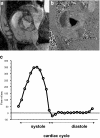
Fig. 1
Fig. 1
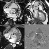
Phase-contrast MRI of an aortic valve in a normal volunteer. Magnitude image (a), phase image (b), corresponding flow map (c). Images a and b show peak of systolic ejection: three-leaflets valve and presence of normal orifice are well visible on magnitude image (a). Phase image (b) shows forward blood flow (shown as a dark area) through opened aortic valve. Stationary tissues are shown as gray. Flow map (c) obtained by delineating the aortic valve on all cardiac time frames, shows a peak systolic forward flow (77 ml/heart beat) and absent significant reversal diastolic flow
Fig. 2
Fig. 2
Eccentric mitral regurgitation. Transesophageal echocardiography (a) shows eccentric jet (arrows) directed from left ventricle (LV) into left atrium (LA) (movie 2A). The jet impinges on LA wall (“wall jet”) dissipating part of its kinetic energy (“jet momentum”), resulting in underestimation of mitral regurgitation severity. Contrast ventriculography (b), obtained in right anterior oblique view, confirms eccentric regurgitant jet (*) (arrows) (movie 2B). Balanced SSFP cine-MRI in LV inflow-outflow view (c) clearly shows eccentric mitral regurgitation (arrows) (movie 2C). Valve defect was not seen on vertical long and horizontal long axis views (not shown), emphasizing the importance of multiplane scanning in eccentric regurgitation
Fig. 3a–d
Fig. 3a–d
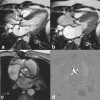
Severe aortic regurgitation. Bicuspid aortic valve (latero-lateral rim) with rupture of anterior leaflet and severe aortic regurgitation with left ventricular (LV) volume overload. Cine MRI (SSFP sequence) perpendicularly oriented through aortic valve (a), along LV outflow tract (b). Magnitude (c) and phase-contrast (d) MR image perpendicular through LV outflow tract. In a, closure of bicuspid aortic valve (arrowhead) but presence of area of signal void (arrow) in middle of anterior leaflet corresponding to the area of rupture. In b, anterior leaflet rupture leads to complex and important holodiastolic aortic regurgitation (movie 3B). Severity of aortic insufficiency is quantified using phase-contrast imaging (movie 3C), where regurgitation is depicted as a pinpoint dark area (c, d; arrows). Following parameters are derived: regurgitation flow volume: 77 ml; regurgitation fraction: 46%. Chronic volume overload has led to important eccentric LV hypertrophy (end-diastolic volume: 328 ml; ejection fraction: 58%; mass: 195 g)
Fig. 4
Fig. 4
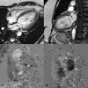
Functional mitral regurgitation in a 44-year-old woman with idiopathic dilated cardiomyopathy. Functional mitral regurgitation is caused by dilation of mitral ring (loss of sphincter-like function) and tethering of both leaflets due to papillary muscles displacement. Cine MRI (SSFP sequence) in horizontal long-axis (a) and vertical long-axis (b), both at systole. Regurgitant flow appears as stripe-like jet (arrows) into left atrium. Phase-contrast MRI (see movie 4) obtained perpendicularly through the mitral valve. During systole (c), regurgitant flow appears as a pinpoint white spot (black arrows), whereas during diastole (d) the inflow through mitral valve is shown as an oval black structure (white arrows). A regurgitation fraction of 12% was derived
Fig. 5
Fig. 5
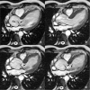
Aortic regurgitation due to annulo-aortic ectasia. A 52-year-old man affected by annulo-aortic ectasia (ascending aorta diameter: 50 mm). Four frames of cine MRI (SSFP sequence) of left ventricular inflow-outflow view: end-diastole (a), mid systole (b), early diastole (c) and mid diastole (d). Stretching of normal aortic valve cusps (not shown) because of aortic root dilatation with consequent loss of coaptation resulting in eccentric valve regurgitation. Regurgitant jet is sharply defined in its proximal portion, where it abuts the anterior mitral leaflet, and then fans out (arrows) into the LV (d; movie 5). Regurgitation fraction is 30% with LV overload (end-diastolic volume: 257 ml; ejection fraction: 52%)
Fig. 6
Fig. 6
Severe pulmonary regurgitation post tetralogy of Fallot repair in a 20-year-old man. SSFP cine MRI (a) of the right ventricle (RV), RV out-flow tract and main pulmonary artery (PA) obtained at diastole. Abnormal coaptation of pulmonary cusps (arrowheads) results in severe pulmonary regurgitation (arrows). Phase image perpendicular to RV outflow tract during systole (b) indicates forward blood flow (“bright” appearance) across outflow tract during systole (arrows). Corresponding phase image during diastole (c) shows severe backward flow (“black” appearance), resulting in regurgitation fraction of 46%, and reduced RV ejection fraction of 43% (d forward flow is shown in green, severe regurgitant diastolic flow in red)
Fig. 7
Fig. 7
Bicuspid aortic valve in a 40-year-old man with prevalent stenosis. Bicuspid aortic valve, type antero-posterior rim, is well seen on magnitude (b) and phase images (c) (arrows). Valve abnormality results in stenosis (peak velocity 4.2 m/s; peak gradient: 70 mmHg, and planimetry aortic orifice: 1.1 cm2), although moderate aortic regurgitation is also present (not shown; regurgitation fraction of 30%). Aortic valve abnormalities have led to severe left ventricular remodeling (end-diastolic volume: 400 ml; ejection fraction 46.5%). Balanced-SSFP cine-MRI (a) during systolic ejection phase shows the “doming sign” (arrowhead) of left cusp and signal void in ascending aorta due to turbulent blood flow (arrows). Cusp doming is very often detected in congenital aortic valve stenosis as valve leaflet mobility is partially preserved due to lack of extensive calcification
Fig. 8
Fig. 8
Recurrence of severe aortic stenosis in a 38-year-old man. He had a history of rheumatic disease, with valvulotomy for aortic stenosis at age of 18. Balanced SSFP cine MRI of left ventricular inflow/outflow view during mid-systole (a) and early diastole (b) (movies 8A, 8B). Signal voids (white arrows) due to turbulent blood flow, shows concomitant presence of stenosis and regurgitation. Phase-contrast MRI, magnitude image (c) and phase image (d), reveals predominant stenotic defect (arrowheads) with peak systolic gradient of 170 mmHg and planimetric aortic valve area of 0.7 cm2, and less severe aortic regurgitation (22%). The aortic valve, is functionally bicuspid due to fusion of two aortic cusps which are extensively calcified (low signal intensity on leaflet tips; see movies). Note presence of dilated ascending aorta (48 mm)
Fig. 9
Fig. 9
Metallic aortic valve prosthesis in a 63-year-old man after Bentall surgery for ascending aortic dissection. Valve prothesis is visible as an area of signal void (white arrow) minimally disturbing image quality. Presence of periprosthetic leak (arrowhead)

Similar articles

Cited by

References

    1. Simpson IA, Sahn DJ. Quantification of valvular regurgitation by Doppler echocardiography. Circulation. 1991;84(3 Suppl):I188–I192. - PubMed
    1. Segal J, Lerner DJ, Miller DC, Mitchell RS, Alderman EA, Popp RL. When should Doppler-determined valve area be better than the Gorlin formula?: Variation in hydraulic constants in low flow states. J Am Coll Cardiol. 1987;9:1294–1305. - PubMed
    1. Krombach GA, Kuhl H, Bucker A, Mahnken AH, Spuntrup E, Lipke C, Schroder J, Gunther RW. Cine MR imaging of heart valve dysfunction with segmented true fast imaging with steady state free precession. J Magn Reson Imaging. 2004;19:59–67. doi: 10.1002/jmri.10428. - DOI - PubMed
    1. Nikolaou K, Flohr T, Knez A, Rist C, Wintersperger B, Johnson T, Reiser MF, Becker CR. Advances in cardiac CT imaging: 64-slice scanner. Int J Cardiovasc Imaging. 2004;20:535–540. doi: 10.1007/s10554-004-7015-1. - DOI - PubMed
    1. Johnson TR, Nikolaou K, Wintersperger BJ, Leber AW, Ziegler F, Rist C, Buhmann S, Knez A, Reiser MF, Becker CR. Dual-source CT cardiac imaging: initial experience. Eur Radiol. 2006;16:1409–1415. doi: 10.1007/s00330-006-0298-y. - DOI - PubMed

Publication types

MeSH terms