. 2008 Apr;57(4):531-9.
doi: 10.1007/s00262-007-0391-3. Epub 2007 Sep 1.

Vgamma9Vdelta2 T cell-mediated recognition of human solid tumors. Potential for immunotherapy of hepatocellular and colorectal carcinomas

Affiliations

Vgamma9Vdelta2 T cell-mediated recognition of human solid tumors. Potential for immunotherapy of hepatocellular and colorectal carcinomas

Francoise Bouet-Toussaint et al. Cancer Immunol Immunother. 2008 Apr.

Abstract

Introduction: Vgamma9Vdelta2 T lymphocytes are reported to participate in the anti-tumor immune surveillance in human. They are known to recognize phosphoantigens and molecules expressed on cells undergoing neoplasic transformation. In this study, we investigated phenotype and anti-tumor cytotoxicity of ex vivo expanded Vgamma9Vdelta2 T cells in view of adoptive immunotherapy.

Materials and methods: Experiments were performed with peripheral blood samples from eleven patients [six colorectal carcinoma (CRC), four hepatocellular carcinoma (HCC), one sarcoma] and sixteen healthy donors.

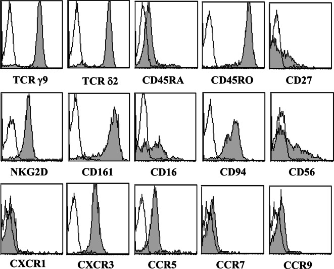
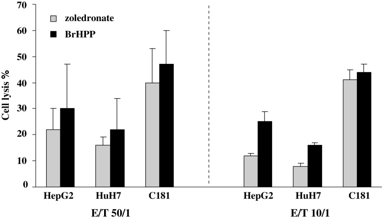
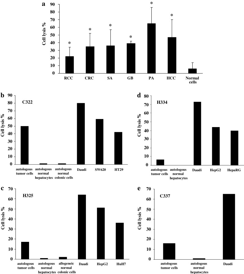
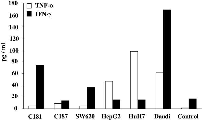
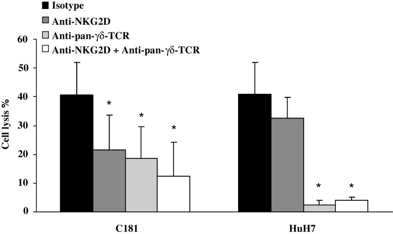
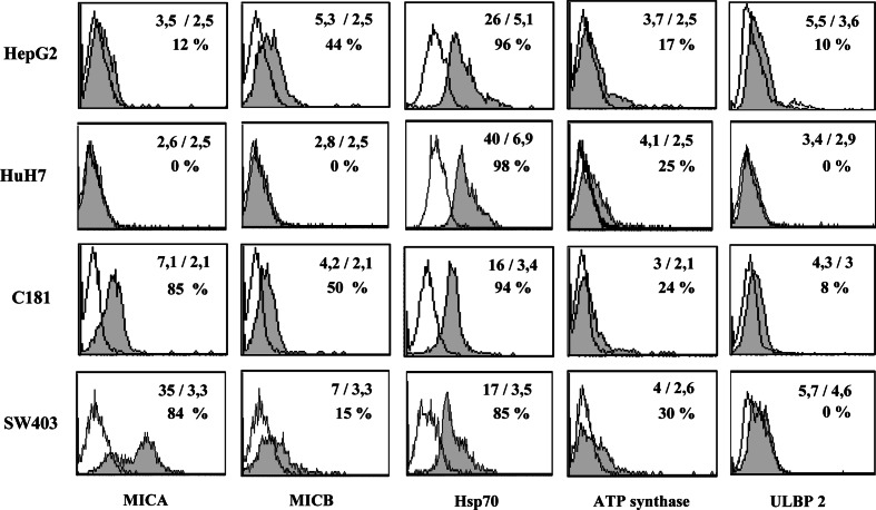
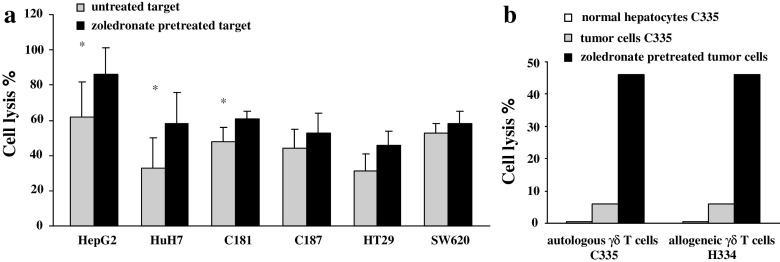
Results/discussion: Ex vivo expansion of Vgamma9Vdelta2 T cells could be achieved by a single dose of phosphoantigen, either bromohydrin pyrophosphate or zoledronate, and supported by exogenous IL-2. After 2 weeks, expanded Vgamma9Vdelta2 T lymphocytes acquired the effector memory phenotype CD45RA(-)CD45RO(high)CD27(-). They expressed NKG2D and CD161 and the proinflammatory CXCR3 and CCR5 chemokine receptors. Vgamma9Vdelta2 T cells displayed a strong lytic activity toward a broad panel of tumor cell lines or primary cultures. Interestingly, HCC and CRC primary cells could be lysed by autologous Vgamma9Vdelta2 T cells whereas autologous normal cells were not sensitive to the lysis. mAbs blocking assays demonstrated that TCR was the most important receptor involved in the lysis of tumor cells. However, NKG2D receptor could deliver a costimulatory signal enhancing the lysis of HCC and CRC tumors expressing MICA/B. Treatment of tumor cells by the mevalonate pathway inhibitor, zoledronate, enhanced the killing of both HCC and CRC. Expansion index of Vgamma9Vdelta2 T cells was in similar levels in healthy donors or in cancer patients and total expansion was suitable for adoptive immunotherapy.

Conclusion: These results provide a rationale for the clinical evaluation of Vgamma9Vdelta2 T lymphocytes in HCC and CRC.


Figures

Fig. 1
Fig. 1
Phenotype of BrHPP expanded Vγ9Vδ2 T cells. PBMCs from donors were treated at day 0 with 300 nM BrHPP and cultured in presence of 400 IU IL-2/ml. The cytometric analysis was performed on cells collected after 17 days of culture using mAbs against γ9-TCR, δ2-TCR, CD16, CD27, CD45RA, CD45RO, CD56, CD94, CD161, NKG2D, CXCR1, CXCR3, CCR5, CCR7, and CCR9. The open histograms represent isotype controls and the filled ones are specific stainings. Data are representative of assays performed with Vγ9Vδ2 T cells expanded with BrHPP from either donor or patient PBMCs
Fig. 2
Fig. 2
Cytotoxic activity of BrHPP and zoledronate expanded Vγ9Vδ2 T cells against tumor cells. PBMCs from three donors were treated at day 0 with 300 nM BrHPP or 1 μM zoledronate and cultured in presence of 400 IU IL-2/ml within 2 weeks. Expanded cells were incubated for 4 h with 5 × 103 target cells previously labeled with 51Cr in ratio effector to target (E/T) of 50/1 and 10/1. Target cells were HepG2 (n = 9) and HuH7 (n = 6) HCC cell lines and C181 (n = 3) CRC cell line
Fig. 3
Fig. 3
Cytotoxic activity of Vγ9Vδ2 T cells from cancer patients against tumor cells from different origins. PBMCs were treated at day 0 with 300 nM BrHPP and cultured in presence of 400 IU IL-2/ml within 2 weeks. Expanded Vγ9Vδ2 T cells were incubated for 4 h with 5 × 103 target cells previously labeled with 51Cr in a ratio effector to target (E/T) of 50/1. Results are the mean of assays performed in triplicate. *: significant difference when compared with cytotoxicity against normal target cells. a Cytotoxic activity in allogeneic context. PBMCs were from seven patients suffering from CRC, HCC or sarcoma. Target cells were RCC (R104, R119, R131 cell lines and R180, R305 primary cultures) (n = 8, P = 0.01), CRC (C181, C187, SW620, SW403, HT29 cell lines, and C293 primary culture) (n = 22, P = 0.001), HCC (HepG2, HuH7, and BC2 cell lines) (n = 8, P = 0.002), sarcoma (SA297, SA309 primary cultures) (n = 4, P = 0.048), glioblastoma (GB2, GB3 primary cultures) (n = 4, P = 0.004), and tumoral pancreatic cells (PA287 primary cultures) (n = 2, P = 0.044). Normal cells used as control targets were normal colonic (n = 2) and normal hepatic cells (n = 2) from patients and PBMCs from healthy donors (n = 8). b–e Selective cytotoxic activity against autologous tumor cells. Target cells were autologous normal and tumor cells isolated from liver or colonic biopsies from patient C322 (b), H325 (c), H334 (d), and C337 (e). Cytotoxicity against normal allogeneic cells and CRC (SW620, HT29), HCC (HepG2, HuH7, and HepaRG), and Daudi cell lines was also evaluated
Fig. 4
Fig. 4
TNF-α and IFN-γ release by Vγ9Vδ2 T cells. PBMCs from two patients (1 CRC and 1 HCC) and two donors were treated at day 0 with 300 nM BrHPP and cultured in presence of 400 IU IL-2/ml within 2 weeks. About 3 × 105 expanded Vγ9Vδ2 T cells were co-cultured with 105 cells from CRC (C181, C187, and SW620), HCC (HepG2, HuH7) and Daudi cell line in 2 ml of complete medium. TNF-α and IFN-γ release was measured, respectively, 6 and 48 h after stimulation in culture supernatants by ELISA. Control was medium alone. Representative data obtained with patient C322 are shown
Fig. 5
Fig. 5
Modulation of the cytotoxic activity of Vγ9Vδ2 T cells by blocking the NKG2D and TCR interactions. PBMCs from two donors were treated at day 0 with 300 nM BrHPP and cultured in presence of 400 IU IL-2/ml within 2 weeks. Expanded Vγ9Vδ2 T cells were incubated with mAb anti-NKG2D or/and anti-pan-γδ-TCR and then co-cultured for 4 h with 5 × 103 cells from cell lines previously labeled with 51Cr at an effector to target (E/T) ratio of 50/1. Data are mean ± SD of experiments with C181 (n = 6) and HuH7 (n = 6) target cell lines. *, P ≤ 0.03 compared with isotype control
Fig. 6
Fig. 6
Phenotype of CRC and HCC tumor cell lines. Cytometric analyses were performed on tumor cell lines using mAbs against MICA, MICB, HSP70, ATP synthase, ULBP1, ULBP2, and ULBP3. Presented data were obtained from two CRC cell lines (C181, SW403) and two HCC cell lines (HepG2, HuH7). They are representative of assays performed with 4 CRC and 2 HCC and 2 RCC tumor cell lines. The open histograms represent isotype controls and the filled ones are specific stainings. Mean of fluorescence (specific staining/isotype) and percentage of positive cells are noted on each histogram
Fig. 7
Fig. 7
Lysis by Vγ9Vδ2 T cells of zoledronate-pretreated tumor cells. Target cells were pretreated overnight with 5 μM zoledronate. Viability of the cells was unchanged after this pretreatment. Then, 5 × 103 target cells were labeled with 51Cr and incubated for 4 h with Vγ9Vδ2 T cells in a ratio effector to target (E/T) of 50/1. a Target cells were from HCC (HepG2, n = 8; HuH7, n = 8) and CRC (C181, n = 10; C187, n = 8; HT29, n = 6; SW620, n = 6). Effector cells were Vγ9Vδ2 T cells expanded from two donors and one CRC patient. *, P ≤ 0.04 compared with untreated target cells. b Target cells were normal hepatic cells and tumor cells isolated from a liver metastasis from the patient C335. Effector cells were autologous (C335) or allogeneic (H334) Vγ9Vδ2 T cells expanded from patients C335 and H334

References

    1. Aldemir H, Prod’homme V, Dumaurier MJ, Retiere C, Poupon G, Cazareth J, Bihl F, Braud VM. Cutting edge: lectin-like transcript 1 is a ligand for the CD161 receptor. J Immunol. 2005;175:7791–7795. - PubMed
    1. Barkholt L, Danielsson R, Calissendorff B, Svensson L, Malihi R, Remberger M, Uzunel M, Thorne A, Ringden O. Indium-111-labelled donor-lymphocyte infusion by way of hepatic artery and radio-frequency ablation against liver metastases of renal and colon carcinoma after allogeneic hematopoietic stem-cell transplantation. Transplantation. 2004;78:697–703. doi: 10.1097/01.TP.0000129807.53523.97. - DOI - PubMed
    1. Battistini L, Caccamo N, Borsellino G, Meraviglia S, Angelini DF, Dieli F, Cencioni MT, Salerno A. Homing and memory patterns of human gammadelta T cells in physiopathological situations. Microbes Infect. 2005;7:510–517. doi: 10.1016/j.micinf.2004.12.008. - DOI - PubMed
    1. Blattman JN, Greenberg PD. Cancer immunotherapy: a treatment for the masses. Science. 2004;305:200–205. doi: 10.1126/science.1100369. - DOI - PubMed
    1. Bonneville M, Fournie JJ. Sensing cell stress and transformation through Vgamma9Vdelta2 T cell-mediated recognition of the isoprenoid pathway metabolites. Microbes Infect. 2005;7:503–509. doi: 10.1016/j.micinf.2005.03.004. - DOI - PubMed

Publication types

MeSH terms

Substances