Comparative Study
. 2007 Aug 31:7:168.
doi: 10.1186/1471-2407-7-168.

High resolution melting for mutation scanning of TP53 exons 5-8

Affiliations
Comparative Study

High resolution melting for mutation scanning of TP53 exons 5-8

Michael Krypuy et al. BMC Cancer. .

Abstract

Background: p53 is commonly inactivated by mutations in the DNA-binding domain in a wide range of cancers. As mutant p53 often influences response to therapy, effective and rapid methods to scan for mutations in TP53 are likely to be of clinical value. We therefore evaluated the use of high resolution melting (HRM) as a rapid mutation scanning tool for TP53 in tumour samples.

Methods: We designed PCR amplicons for HRM mutation scanning of TP53 exons 5 to 8 and tested them with DNA from cell lines hemizygous or homozygous for known mutations. We assessed the sensitivity of each PCR amplicon using dilutions of cell line DNA in normal wild-type DNA. We then performed a blinded assessment on ovarian tumour DNA samples that had been previously sequenced for mutations in TP53 to assess the sensitivity and positive predictive value of the HRM technique. We also performed HRM analysis on breast tumour DNA samples with unknown TP53 mutation status.

Results: One cell line mutation was not readily observed when exon 5 was amplified. As exon 5 contained multiple melting domains, we divided the exon into two amplicons for further screening. Sequence changes were also introduced into some of the primers to improve the melting characteristics of the amplicon. Aberrant HRM curves indicative of TP53 mutations were observed for each of the samples in the ovarian tumour DNA panel. Comparison of the HRM results with the sequencing results revealed that each mutation was detected by HRM in the correct exon. For the breast tumour panel, we detected seven aberrant melt profiles by HRM and subsequent sequencing confirmed the presence of these and no other mutations in the predicted exons.

Conclusion: HRM is an effective technique for simple and rapid scanning of TP53 mutations that can markedly reduce the amount of sequencing required in mutational studies of TP53.


Figures

Figure 1
Figure 1
Schematic representation of amplicons for the TP53 HRM assay. Forward primers are in blue and reverse are in red. Exons 5 to 8 are represented by the green bars and the amplicon length and name are listed in the lower panels. Two amplicons were designed to span TP53 exon 5.
Figure 2
Figure 2
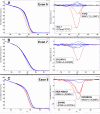
TP53 cell line mutations detected by HRM. Cell line mutant samples are in red and wild-type samples are in blue. Normalised plots are on the left and the corresponding difference plots are on the right. Panel A - T47D 580C>T mutation in exon 6. Panel B - OVCAR-3 743G>A mutation in exon 7. Panel C - MDA-MB231 839G>A, SW480 818G>A, RPMI8226 853G>A mutations in exon 8.
Figure 3
Figure 3
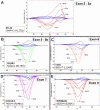
TP53 exon 5 amplicon redesign allowed detection of the BT-20 645A>C mutation. Cell line mutant samples are in red and wild-type samples are in blue. All plots shown in this figure are difference plots. Panel A; BT-20 394A>C and SKBR3 524G>A mutations with the original amplicon designed for exon 5. Panel B; BT-20 394A>C mutation is now detectable with the 5a amplicon. Panel C; SKBR3 524G>A mutation with the 5b amplicon.
Figure 4
Figure 4
Dilution of cell line mutant DNA to test sensitivity of TP53 HRM amplicons. Wild-type samples are in blue and all plots shown are difference plots. Panel A: BT-20 dilution series. Panel B; SKBR3 dilution series. Panel C; T47D dilution series. Panel D; OVCAR-3 dilution series. Panel E; RPMI8226 dilution series.
Figure 5
Figure 5
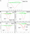
Example of aberrant melt profiles observed for ovarian tumour samples. Wild-type control samples are in blue. Wild-type patient samples are in green and mutant patient samples are in red. Panel A; mutation in patient 33 in exon 5 with amplicon 5a. Panel B; mutations in patients 9, 13 and 14 in exon 5 with amplicon 5b. Panel C; mutation in patient 4 in exon 6. Panel D; mutations in patients 21 and 28 in exon 7. Panel E; mutations in patients 6, 15 and 17 in exon 8.
Figure 6
Figure 6
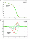
Subtle change in melt profile observed for patient 22 in TP53 exon 6. Wild-type control samples are in blue. Wild-type patient samples are in green and mutant patient samples are in red. Panel A; normalised plot of patient 22 compared to wild-type controls and wild-type ovarian tumour samples in exon 6. Panel B; difference plot of patient 22 compared to wild-type controls and wild-type ovarian tumour samples.
Figure 7
Figure 7
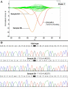
HRM difference plot and sequencing data for breast tumour samples for TP53 exon 7. Panel A; Difference plot of breast tumour samples for TP53 exon 7. Wild-type control samples are in blue, wild-type patient samples are in green, the cell line OVCAR-3 is in red and samples B4 and B9 are in orange. Panel B; Sequencing traces for wild-type, B4 and B9.
Figure 8
Figure 8
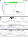
HRM difference plot and sequencing data for breast tumour samples for TP53 exon 5, 5b amplicon. Panel A; Difference plot of breast tumour samples for TP53 exon 5, amplicon 5b. Wild-type control samples are in blue, wild-type patient samples are in green, the cell line SKBR3 is in red, sample B6 is in orange and sample B14 is in pink. Panel B; Sequencing traces for wild-type, B6 and B14.

Similar articles

Cited by

References

    1. Diller L, Kassel J, Nelson CE, Gryka MA, Litwak G, Gebhardt M, Bressac B, Ozturk M, Baker SJ, Vogelstein B, et al. p53 functions as a cell cycle control protein in osteosarcomas. Mol Cell Biol. 1990;10:5772–5781. - PMC - PubMed
    1. Deppert W, Buschhausen-Denker G, Patschinsky T, Steinmeyer K. Cell cycle control of p53 in normal (3T3) and chemically transformed (Meth A) mouse cells. II. Requirement for cell cycle progression. Oncogene. 1990;5:1701–1706. - PubMed
    1. Lin D, Shields MT, Ullrich SJ, Appella E, Mercer WE. Growth arrest induced by wild-type p53 protein blocks cells prior to or near the restriction point in late G1 phase. Proc Natl Acad Sci U S A. 1992;89:9210–9214. doi: 10.1073/pnas.89.19.9210. - DOI - PMC - PubMed
    1. Yonish-Rouach E, Grunwald D, Wilder S, Kimchi A, May E, Lawrence JJ, May P, Oren M. p53-mediated cell death: relationship to cell cycle control. Mol Cell Biol. 1993;13:1415–1423. - PMC - PubMed
    1. Kuerbitz SJ, Plunkett BS, Walsh WV, Kastan MB. Wild-type p53 is a cell cycle checkpoint determinant following irradiation. Proc Natl Acad Sci U S A. 1992;89:7491–7495. doi: 10.1073/pnas.89.16.7491. - DOI - PMC - PubMed

Publication types

Substances