

The role of proopiomelanocortin (POMC) neurones in feeding behaviour
- PMID: 17764572
- PMCID: PMC2018708
- DOI: 10.1186/1743-7075-4-18
The role of proopiomelanocortin (POMC) neurones in feeding behaviour
Abstract
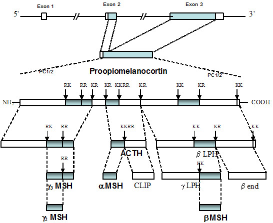
The precursor protein, proopiomelanocortin (POMC), produces many biologically active peptides via a series of enzymatic steps in a tissue-specific manner, yielding the melanocyte-stimulating hormones (MSHs), corticotrophin (ACTH) and beta-endorphin. The MSHs and ACTH bind to the extracellular G-protein coupled melanocortin receptors (MCRs) of which there are five subtypes. The MC3R and MC4R show widespread expression in the central nervous system (CNS), whilst there is low level expression of MC1R and MC5R. In the CNS, cell bodies for POMC are mainly located in the arcuate nucleus of the hypothalamus and the nucleus tractus solitarius of the brainstem. Both of these areas have well defined functions relating to appetite and food intake. Mouse knockouts (ko) for pomc, mc4r and mc3r all show an obese phenotype, as do humans expressing mutations of POMC and MC4R. Recently, human subjects with specific mutations in beta-MSH have been found to be obese too, as have mice with engineered beta-endorphin deficiency. The CNS POMC system has other functions, including regulation of sexual behaviour, lactation, the reproductive cycle and possibly central cardiovascular control. However, this review will focus on feeding behaviour and link it in with the neuroanatomy of the POMC neurones in the hypothalamus and brainstem.
Figures
References
-
- Sawchenko PE, Brown ER, Chan RKW, Ericsson A, Li H-Y, Roland BL, Kovács KJ. The paraventricular nucleus of the hypothalamus and the functional neuroanatomy of visceromotor responses to stress. Prog Brain Res. 1996;107:201–222. - PubMed
-
- Millington GWM, Buckingham JC. Thymic peptides and neuroendocrine immune communication. J Endocrinol. 1992;133:163–168. - PubMed
LinkOut - more resources
Full Text Sources
Research Materials
Miscellaneous
