. 2007 Oct 1;310(1):154-68.
doi: 10.1016/j.ydbio.2007.07.036. Epub 2007 Aug 3.

Hand transcription factors cooperatively regulate development of the distal midline mesenchyme

Affiliations

Hand transcription factors cooperatively regulate development of the distal midline mesenchyme

Ana C Barbosa et al. Dev Biol. .

Abstract

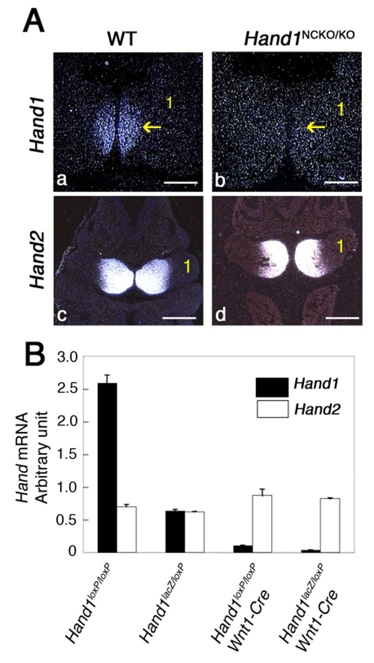
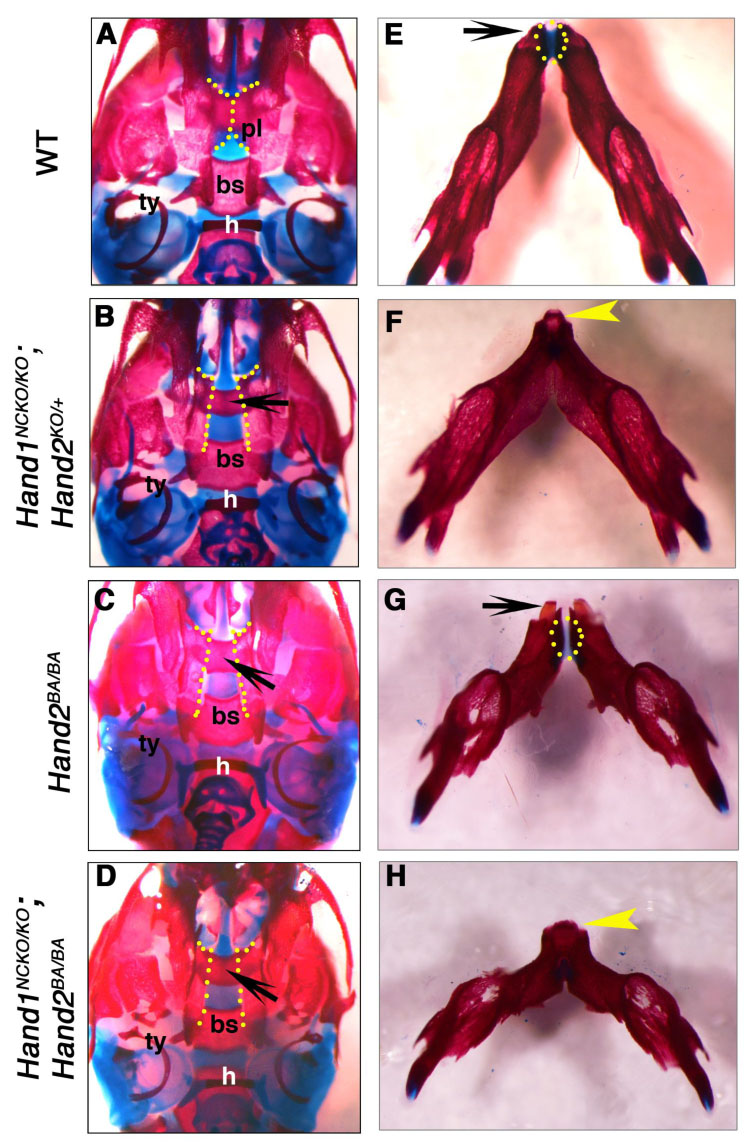
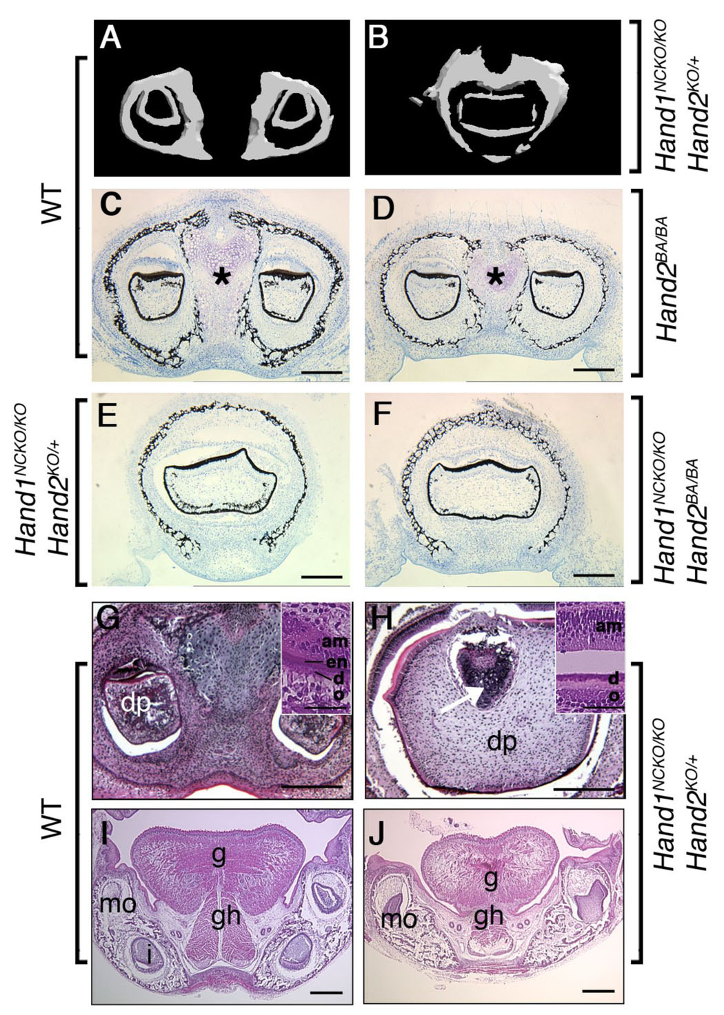
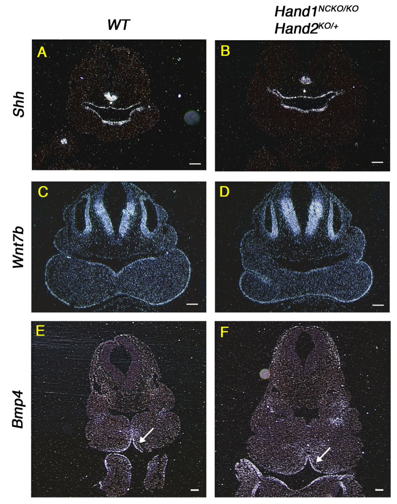
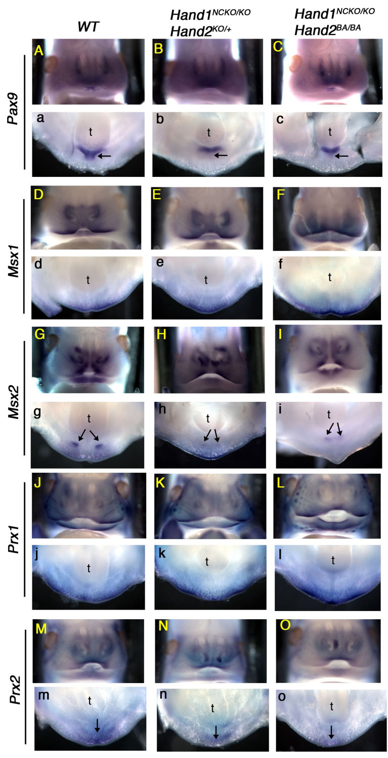
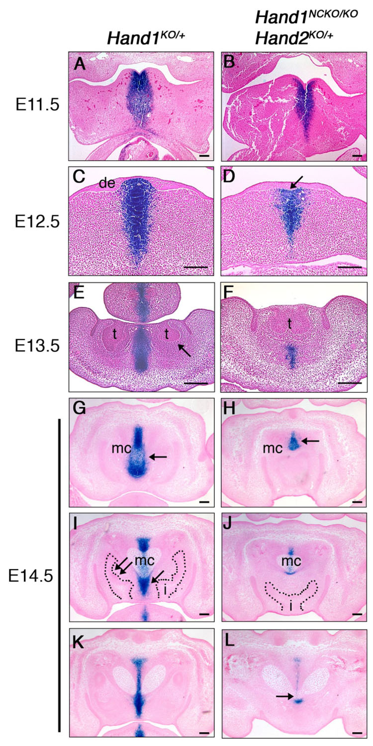
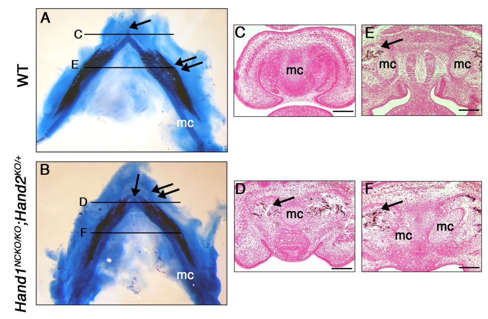
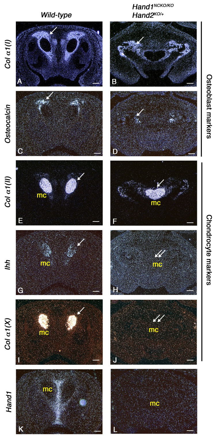
Hand proteins are evolutionally conserved basic helix-loop-helix (bHLH) transcription factors implicated in development of neural crest-derived tissues, heart and limb. Hand1 is expressed in the distal (ventral) zone of the branchial arches, whereas the Hand2 expression domain extends ventrolaterally to occupy two-thirds of the mandibular arch. To circumvent the early embryonic lethality of Hand1 or Hand2-null embryos and to examine their roles in neural crest development, we generated mice with neural crest-specific deletion of Hand1 and various combinations of mutant alleles of Hand2. Ablation of Hand1 alone in neural crest cells did not affect embryonic development, however, further removing one Hand2 allele or deleting the ventrolateral branchial arch expression of Hand2 led to a novel phenotype presumably due to impaired growth of the distal midline mesenchyme. Although we failed to detect changes in proliferation or apoptosis between the distal mandibular arch of wild-type and Hand1/Hand2 compound mutants at embryonic day (E)10.5, dysregulation of Pax9, Msx2 and Prx2 was observed in the distal mesenchyme at E12.5. In addition, the inter-dental mesenchyme and distal symphysis of Meckel's cartilage became hypoplastic, resulting in the formation of a single fused lower incisor within the hypoplastic fused mandible. These findings demonstrate the importance of Hand transcription factors in the transcriptional circuitry of craniofacial and tooth development.


Figures

Figure 1
Figure 1. Neural crest-specific Cre-mediated recombination of Hand1
(A). Expression of Hand1 and Hand2 was examined by in situ hybridization of transverse sections of E10.5 wild-type (panels a, c) and Hand1NCKO/KO (panels b, d) embryos. Hand1 expression seen in the distal part of the mandibular arch (panel a, arrow) is absent in the mutant (panel b, arrow); Hand2 expression is unaffected (panel d). Bars indicate 200 µm (panels a, b) and 400 µm (panels c, d). 1; first mandibular arch. (B). Detection of Hand1 and Hand2 by real-time RT-PCR using RNA extracted from mandibular and second branchial arches of Hand1loxP/loxP, Hand1lacZ/loxp and Hand1NCKO/KO (Hand1loxP/loxP; Wnt1::Cre and Hand1lacZ/loxP;Wnt1::Cre) embryos at E10.5. Values are normalized to 18S expression. Hand1 expression is almost undetectable in the mutants, whereas Hand2 expression is maintained in embryos from all genotypes. Bars are means ± SD.
Figure 2
Figure 2. Craniofacial defects in P1 mice with compound mutations in Hand1 and Hand2
Bone and cartilage staining of wild-type (A, E), Hand1NCKO; Hand2KO/+ (B, F), Hand2BA/BA (C, G) and Hand1NCKO; Hand2BA/BA (D, H) mutant mice are shown. (A–D) Ventral views of the skull. Fusion of the bilateral palatine processes observed in the wild-type mouse (A, dashed line) is absent in the mutants (B-D, dashed line). Underlying presphenoid bone is visible in the mutants (arrows in B–D). (E–H) Ventral views of the mandible. The mandible of the mutants (F–G) is shorter and deformed. The angle between the left and right mandible is wider compared to the wild-type mouse (E). Note that the deformity of the mandible increases as total Hand1 and Hand2 gene dosage decreases. Wild-type (E) and Hand2BA/BA (G) mice show two incisors at the distal tip (arrow), whereas the mandible and lower incisors are fused in Hand1NCKO; Hand2KO/+ (F) and Hand1NCKO; Hand2BA/BA (H) mice (arrowhead). Note that the distal symphysis of the mandible is present in the wild-type and Hand2BA/BA mice (E, G, dotted area) but absent in the compound mutants (F, H).
Figure 3
Figure 3. Abnormal fusion of the lower incisors
(A, B) Micro-CT analysis of the lower incisors in P1 embryos. In wild-type embryo (A), two lower incisors are located within the mandible. In contrast, a large deformed single incisor is observed within the fused mandible of the mutant (B). (C–F) Von Kossa staining of coronal sections of the mandible in plastic-embedded wild-type (C), Hand2BA/BA (D), Hand1NCKO/KO;Hand2KO/+ (E) and Hand1NCKO/KO;Hand2BA/BA (F) embryos. In wild-type embryos, a well-developed distal symphysis of Meckel’s cartilage is observed (asterisk in C). The symphysis is much smaller in the Hand2BA/BA mutant (asterisk in D) and becomes undetectable in Hand1NCKO/KO;Hand2KO/+ and Hand1NCKO/KO;Hand2BA/BA embryos (E, F). Fused incisors are seen in mutant embryos (E, F). (G–J) H&E staining of coronal paraffin sections of P1 wild-type (G, I) and Hand1NCKO/KO;Hand2KO/+ (H, J) embryos. (G, H) In wild-type embryos (G), two incisors with normal cytodifferentiation, including dental papilla (dp), odontoblasts (o), dentin (d), enamel (en) and ameloblasts (am) are observed (inset in G). In the mutant (H), a large fused incisor is observed, and the enamel layer is absent (inset in H). The distal symphysis is embedded in the dental papilla (H, arrow). (I, J) Well-developed extrinsic muscles (g; genioglossus, gh; geniohyoid) are observed in the tongue of wild-type embryos (I). In the mutant (J), muscle fibers are sparse and hypoplastic. Bars indicate 100 µm (C–J) and 40 µm (inset in G and H). mo; molar, i; incisor.
Figure 4
Figure 4. Normal specification of the odontogenic epithelium in E10.5 mutant embryos
In situ hybridization of transverse sections from E10.5 wild-type (A, C, E) and Hand1NCKO/KO;Hand2KO/+ (B, D, F) embryos probed with Shh (A, B), Wnt7b (C, D) and Bmp4 (E, F). Arrow in E and F indicates that a Bmp4 domain is not altered in the mutant embryo compared with wild-type embryo. Bars indicate 100 µm.
Figure 5
Figure 5. Dysregulation of branchial arch genes in embryos with compound mutations in Hand1 and Hand2
Whole-mount in situ hybridization was performed in E12.5 wild-type (A, a, D, d, G, g, J, j, M, m), Hand1NCKO/KO;Hand2KO/+ (B, b, E, e, H, h, K, k, N, n) and Hand1NCKO/KO;Hand2BA/BA (C, c, F, f, I, i, L, l, O, o) embryos. Frontal views (upper case) and transverse oral views (lower case) are shown. Pax9 is expressed in the dental and inter-dental (arrow in a) mesenchyme of the wild-type embryos, however, inter-dental expression is absent in both mutant embryos (arrows in b, c). Msx2 is expressed in the incisors (arrows in g) and Prx2 is expressed in the distal midline mesenchyme of wild-type embryos (arrow in m). In both mutants, Msx2 and Prx2 are down-regulated (arrows in h, i, arrow in n, o). Other candidate marker genes, Msx1 and Prx1, showed no remarkable changes.
Figure 6
Figure 6. Development of Hand1-positive distal midline mesenchyme during embryogenesis
LacZ staining of Hand1KO/+ (A, C, E, G, I, K) and Hand1NCKO/KO;Hand2KO/+ (B, D, F, H, J, L) embryos. Transverse sections (A–F) and coronal sections (G–L) are shown. (A, B) LacZ staining is observed in the distal tip of the mandibular arch in both wild-type and mutant embryos at E11.5. (C, D) At E12.5, two distinct thickenings of the dental epithelium (de) corresponding to the future incisors are observed in wild-type embryo (C), whereas one linear thickening of the epithelium was observed in the mutant (D, arrow). (E, F) In wild-type embryos, two well-developed incisor buds are observed. Strong lacZ staining was observed in the distal midline mesenchyme between the tooth buds and in the center of the tongue in wild-type embryo (E). Dental mesenchyme is negative for lacZ (E, arrow). In the mutant, lacZ-positive mesenchyme is markedly reduced and tooth buds are fused (F). (G–L) Sections were taken in a distal (G, H) to proximal (K, L) direction in E14.5 embryos. In wild-type embryos, strong lacZ staining is detected in the distal symphysis of the Meckel’s cartilage (G, arrow) and the inter-dental mesenchyme (I, arrow). Two cap-stage incisors are observed (i in I). Dental mesenchyme is negative for lacZ (I, double arrow). In the mutant, the distal symphysis is significantly reduced in size (H, arrow), and a single incisor is observed (i in J). The distal end of Meckel’s cartilage is tapered and fused (arrow in L). t; tooth bud, i; incisor, mc; Meckel’s cartilage. Bars indicate 100 µm.
Figure 7
Figure 7. Premature mineralization of the mandible in Hand1NCKO/KO; Hand2KO/+ embryos
(A, B) Alcian blue and alizarin red staining of E14.5 wild-type (A) and Hand1NCKO/KO;Hand2KO/+ (B) embryos. In wild-type embryo (A), a well-developed distal symphysis of the Meckel’s cartilage (arrow) and mineralization of the mandible (double arrow) is observed along the body of Meckel’s cartilage. In the mutant (B), the distal symphysis is absent (arrow) and mineralization is accelerated at the distal portion of Meckel’s cartilage (double arrows). (C–F) Coronal sections of the mandible from E14.5 wild-type (C, E) and mutant (D, F) embryos stained with Von Kossa. Levels are indicated in A and B. Mineralization is observed around the proximal portion of Meckel’s cartilage in both wild-type and mutant embryos (arrow in E and F). In contrast, mineralization in the distal tip of the Meckel’s cartilage is only observed in the mutant (arrow in D, compare C and D). Bars indicate 100 µm. mc; Meckel’s cartilage.
Figure 8
Figure 8. Expression of bone and cartilage differentiation markers in E14.5 wild-type and Hand1NCKO/KO;Hand2KO/+ embryos
Coronal sections of the mandible from wild-type (A, C, E, G, I, K) and mutant embryos (B, D, F, H, J, L) are hybridized with osteoblast differentiation markers (A–D) or chondrocyte differentiation markers (E–J). Membranous bone ossification takes place around Meckel’s cartilage (arrows in A–D). While collagen αI (I) is expressed in both wild-type (A) and mutant (B) embryos, osteocalcin, a mature osteoblast marker, is down-regulated in the mutant (D). Note that Ihh and collagen α1 (X) expressed in the wild-type Meckel’s cartilage (arrows in G, I) are absent in the mutant embryos (double arrows in H, J). Hand1 expression performed as a control is absent in the mutant embryo (K, L). Bars indicate 100 µm.

References

    1. Abe M, Tamamura Y, Yamagishi H, Maeda T, Kato J, Tabata MJ, Srivastava D, Wakisaka S, Kurisu K. Tooth-type specific expression of dHAND/Hand2: possible involvement in murine lower incisor morphogenesis. Cell Tissue Res. 2002;310:201–212. - PubMed
    1. Aigner T, Dietz U, Stoss H, von der Mark K. Differential expression of collagen types I, II, III, and X in human osteophytes. Lab Invest. 1995;73:236–243. - PubMed
    1. Akiyama H, Shigeno C, Iyama K, Ito H, Hiraki Y, Konishi J, Nakamura T. Indian hedgehog in the late-phase differentiation in mouse chondrogenic EC cells, ATDC5: upregulation of type X collagen and osteoprotegerin ligand mRNAs. Biochem Biophys Res Commun. 1999;257:814–820. - PubMed
    1. Chai Y, Jiang X, Ito Y, Bringas P, Jr, Han J, Rowitch DH, Soriano P, McMahon AP, Sucov HM. Fate of the mammalian cranial neural crest during tooth and mandibular morphogenesis. Development. 2000;127:1671–1679. - PubMed
    1. Chai Y, Maxson RE., Jr Recent advances in craniofacial morphogenesis. Dev Dyn. 2006;235:2353–2375. - PubMed

Publication types