

Perirhinal contributions to human visual perception
- PMID: 17764947
- PMCID: PMC1971135
- DOI: 10.1016/j.cub.2007.07.066
Perirhinal contributions to human visual perception
Abstract
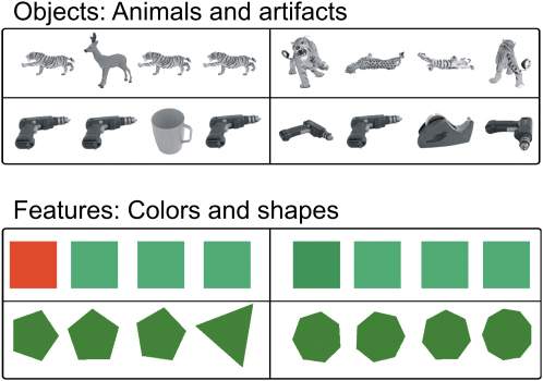
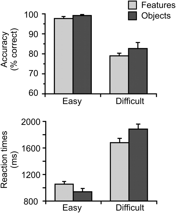
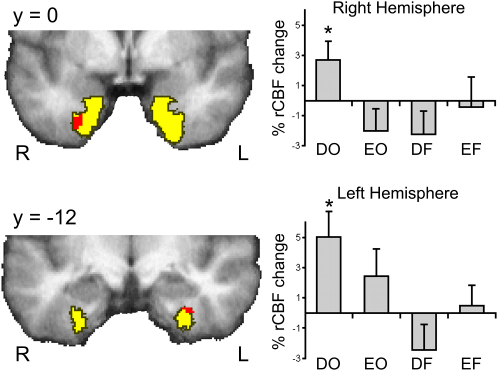
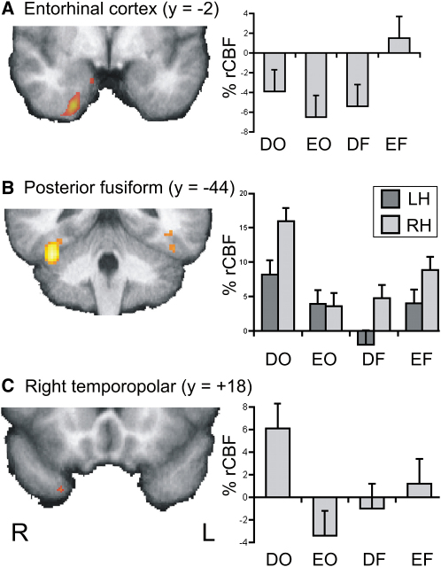
Medial temporal lobe (MTL) structures including the hippocampus, entorhinal cortex, and perirhinal cortex are thought to be part of a unitary system dedicated to memory [1, 2], although recent studies suggest that at least one component-perirhinal cortex-might also contribute to perceptual processing [3, 4, 5, 6]. To date, the strongest evidence for this comes from animal lesion studies [7, 8, 9, 10, 11, 12, 13, 14]. In contrast, the findings from human patients with naturally occurring MTL lesions are less clear and suggest a possible functional difference between species [15, 16, 17, 18, 19, 20]. Here, both these issues were addressed with functional neuroimaging in healthy volunteers performing a perceptual discrimination task originally developed for monkeys [7]. This revealed perirhinal activation when the task required the integration of visual features into a view-invariant representation but not when it could be accomplished on the basis of simple features (e.g., color and shape). This activation pattern matched lateral inferotemporal regions classically associated with visual processing but differed from entorhinal cortex associated with memory encoding. The results demonstrate a specific role for the perirhinal cortex in visual perception and establish a functional homology for perirhinal cortex between species, although we propose that in humans, the region contributes to a wider behavioral repertoire including mnemonic, perceptual, and linguistic processes.
Figures
Similar articles
-
Perirhinal and hippocampal contributions to visual recognition memory can be distinguished from those of occipito-temporal structures based on conscious awareness of prior occurrence.Hippocampus. 2007;17(11):1081-92. doi: 10.1002/hipo.20347. Hippocampus. 2007. PMID: 17696171
-
Involvement of the human medial temporal lobe in a visual discrimination task.Behav Brain Res. 2014 Jul 15;268:22-30. doi: 10.1016/j.bbr.2014.03.030. Epub 2014 Mar 24. Behav Brain Res. 2014. PMID: 24675159
-
Perceptual deficits in amnesia: challenging the medial temporal lobe 'mnemonic' view.Neuropsychologia. 2005;43(1):1-11. doi: 10.1016/j.neuropsychologia.2004.07.017. Neuropsychologia. 2005. PMID: 15488899 Clinical Trial.
-
The perceptual-mnemonic/feature conjunction model of perirhinal cortex function.Q J Exp Psychol B. 2005 Jul-Oct;58(3-4):269-82. doi: 10.1080/02724990544000004. Q J Exp Psychol B. 2005. PMID: 16194969 Review.
-
The role of the perirhinal cortex and hippocampus in learning, memory, and perception.Q J Exp Psychol B. 2005 Jul-Oct;58(3-4):246-68. doi: 10.1080/02724990444000186. Q J Exp Psychol B. 2005. PMID: 16194968 Review.
Cited by
-
Integrative and distinctive coding of visual and conceptual object features in the ventral visual stream.Elife. 2018 Feb 2;7:e31873. doi: 10.7554/eLife.31873. Elife. 2018. PMID: 29393853 Free PMC article.
-
Human medial temporal lobe damage can disrupt the perception of single objects.J Neurosci. 2010 May 12;30(19):6588-94. doi: 10.1523/JNEUROSCI.0116-10.2010. J Neurosci. 2010. PMID: 20463221 Free PMC article.
-
Dissociable roles of the inferior longitudinal fasciculus and fornix in face and place perception.Elife. 2015 Aug 29;4:e07902. doi: 10.7554/eLife.07902. Elife. 2015. PMID: 26319355 Free PMC article.
-
Demands on perceptual and mnemonic fidelity are a key determinant of age-related cognitive decline throughout the lifespan.J Exp Psychol Gen. 2024 Jan;153(1):200-223. doi: 10.1037/xge0001476. J Exp Psychol Gen. 2024. PMID: 38236240 Free PMC article.
-
Age-related impairment in a complex object discrimination task that engages perirhinal cortex.Hippocampus. 2012 Oct;22(10):1978-89. doi: 10.1002/hipo.22069. Hippocampus. 2012. PMID: 22987676 Free PMC article. Clinical Trial.
References
-
- Squire L.R., Stark C.E., Clark R.E. The medial temporal lobe. Annu. Rev. Neurosci. 2004;27:279–306. - PubMed
-
- Murray E.A., Bussey T.J. Perceptual-mnemonic functions of the perirhinal cortex. Trends Cogn. Sci. 1999;3:142–151. - PubMed
-
- Buckley M.J., Gaffan D. Perirhinal cortical contributions to object perception. Trends Cogn. Sci. 2006;10:100–107. - PubMed
-
- Bussey T.J., Saksida L.M. Object memory and perception in the medial temporal lobe: an alternative approach. Curr. Opin. Neurobiol. 2005;15:730–737. - PubMed
Publication types
MeSH terms
Grants and funding
LinkOut - more resources
Full Text Sources
