Structural features of metabolite-sensing riboswitches
- PMID: 17764952
- PMCID: PMC2933830
- DOI: 10.1016/j.tibs.2007.08.005
Structural features of metabolite-sensing riboswitches
Abstract
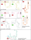
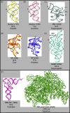
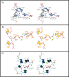
Riboswitches, metabolite-sensing RNA elements that are present in untranslated regions of the transcripts that they regulate, possess extensive tertiary structure to couple metabolite binding to genetic control. Here we discuss recently published structures from four riboswitch classes and compare these natural RNA structures to those of in-vitro-selected RNA aptamers, which bind ligands similar to those of the riboswitches. In addition, we examine the glmS riboswitch - the first example of a ribozyme-based riboswitch. This RNA provides the latest twist in the riboswitch field and portends exciting advances in the coming years. Our knowledge of the mechanisms underlying genetic regulation by riboswitches has increased mightily in recent years and will continue to grow as new riboswitch classes and ligands are discovered and structurally characterized.
Figures


Similar articles
-
[The adenine riboswitch: a new gene regulation mechanism].Med Sci (Paris). 2006 Dec;22(12):1053-9. doi: 10.1051/medsci/200622121053. Med Sci (Paris). 2006. PMID: 17156726 Review. French.
-
Secondary structural entropy in RNA switch (Riboswitch) identification.BMC Bioinformatics. 2015 Apr 28;16:133. doi: 10.1186/s12859-015-0523-2. BMC Bioinformatics. 2015. PMID: 25928324 Free PMC article.
-
Preparation and Crystallization of Riboswitches.Methods Mol Biol. 2016;1320:21-36. doi: 10.1007/978-1-4939-2763-0_3. Methods Mol Biol. 2016. PMID: 26227035
-
Fundamental studies of functional nucleic acids: aptamers, riboswitches, ribozymes and DNAzymes.Chem Soc Rev. 2020 Oct 21;49(20):7331-7353. doi: 10.1039/d0cs00617c. Epub 2020 Sep 18. Chem Soc Rev. 2020. PMID: 32944725 Review.
-
Structural basis of glmS ribozyme activation by glucosamine-6-phosphate.Science. 2006 Sep 22;313(5794):1752-6. doi: 10.1126/science.1129666. Science. 2006. PMID: 16990543
Cited by
-
Analyzing genomic data using tensor-based orthogonal polynomials with application to synthetic RNAs.NAR Genom Bioinform. 2020 Dec 11;2(4):lqaa101. doi: 10.1093/nargab/lqaa101. eCollection 2020 Dec. NAR Genom Bioinform. 2020. PMID: 33575645 Free PMC article.
-
Prediction of hydrogen and carbon chemical shifts from RNA using database mining and support vector regression.J Biomol NMR. 2015 Sep;63(1):39-52. doi: 10.1007/s10858-015-9961-4. Epub 2015 Jul 4. J Biomol NMR. 2015. PMID: 26141454 Free PMC article.
-
Key labeling technologies to tackle sizeable problems in RNA structural biology.Int J Mol Sci. 2008 Jun;9(7):1214-1240. doi: 10.3390/ijms9071214. Epub 2008 Jul 14. Int J Mol Sci. 2008. PMID: 19325801 Free PMC article.
-
Structure of the 30 kDa HIV-1 RNA Dimerization Signal by a Hybrid Cryo-EM, NMR, and Molecular Dynamics Approach.Structure. 2018 Mar 6;26(3):490-498.e3. doi: 10.1016/j.str.2018.01.001. Epub 2018 Feb 2. Structure. 2018. PMID: 29398526 Free PMC article.
-
Tracking RNA with light: selection, structure, and design of fluorescence turn-on RNA aptamers.Q Rev Biophys. 2019 Aug 19;52:e8. doi: 10.1017/S0033583519000064. Q Rev Biophys. 2019. PMID: 31423956 Free PMC article. Review.
References
-
- Hannon GJ, et al. The expanding universe of noncoding RNAs. Cold Spring Harbor Symp. Quant. Biol. 2006;71:551–564. - PubMed
-
- Gollnick P, Babitzke P. Transcription attenuation. Biochim Biophys Acta. 2002;1577:240–250. - PubMed
-
- Winkler WC. Metabolic monitoring by bacterial mRNAs. Arch. Microbiol. 2005;183:151–159. - PubMed
-
- Winkler WC, et al. Control of gene expression by a natural metabolite-responsive ribozyme. Nature. 2004;428:281–286. - PubMed
-
- Brantl S. Regulatory mechanisms employed by cis-encoded antisense RNAs. Curr. Opin. Microbiol. 2007;10:1–8. - PubMed
Publication types
MeSH terms
Substances
Grants and funding
LinkOut - more resources
Full Text Sources

