. 2007 Sep;13(3):433-45.
doi: 10.1016/j.devcel.2007.07.003.

A pathway containing the Ipl1/aurora protein kinase and the spindle midzone protein Ase1 regulates yeast spindle assembly

Affiliations

A pathway containing the Ipl1/aurora protein kinase and the spindle midzone protein Ase1 regulates yeast spindle assembly

Chitra V Kotwaliwale et al. Dev Cell. 2007 Sep.

Abstract

It is critical to elucidate the pathways that mediate spindle assembly and therefore ensure accurate chromosome segregation during cell division. Our studies of a unique allele of the budding yeast Ipl1/Aurora protein kinase revealed that it is required for centrosome-mediated spindle assembly in the absence of the BimC motor protein Cin8. In addition, we found that the Ase1 spindle midzone-associated protein is required for bipolar spindle assembly. The cin8 ipl1 and cin8 ase1 double mutant cells exhibit similar defects, and Ase1 overexpression completely restores spindle assembly in cin8 ipl1 strains. Consistent with the possibility that Ipl1 regulates Ase1, an ase1 mutant lacking the Ipl1 consensus phosphorylation sites cannot assemble spindles in the absence of Cin8. In addition, Ase1 phosphorylation and localization were altered in an ipl1 mutant. We therefore propose that Ipl1/Aurora and Ase1 constitute a previously unidentified spindle assembly pathway that becomes essential in the absence of Cin8.


Figures
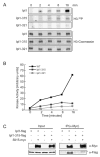
Figure 1
Figure 1
Ipl1–315 is defective in kinase activity and interaction with the Sli15 activator. (A) Ipl1-flag (SBY3672), Ipl1–315-flag (SBY3717), and Ipl1–321-flag (SBY4541) were immunoprecipitated with anti-flag antibody and incubated with histone H3 in a kinase reaction at 30 °C in vitro. The autoradiograms (top three panels) show H3 phosphorylation and the Coomassie-stained gels (lower three panels) show H3 loading. Equal amounts of Ipl1, Ipl1–315, and Ipl1–321 were immunoprecipitated (data not shown). (B) Quantification of (A). (C) Cells expressing either Ipl1-flag (SBY3672), Ipl1-flag and Sli15-myc (SBY5256), Ipl1–315-flag (SBY3717), or Ipl1–315-flag and Sli15-myc (SBY5281) were immunoprecipitated with anti-myc antibody. The extracts (input) and immunoprecipitates (IP) were analyzed by anti-myc (top) and anti-flag (bottom) immunoblotting. Ipl1-flag migrates just below a cross-reacting band (*) in the IP lanes.
Figure 2
Figure 2
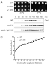
Ipl1–315 is proficient in chromosome segregation, the spindle checkpoint and spindle disassembly. (A) Five-fold serial dilutions of wild-type (SBY3), ipl1–321 (SBY2189), and ipl1–315 (SBY1315) cells were plated at 23 and 37 °C. (B) Wild-type (SBY818), mcd1-1 (SBY870), and mcd1-1 ipl1–315 (SBY1379) cells containing Pds1-myc were arrested in G1 and released to 37 °C. Lysates were prepared at the indicated time points and immunoblotted with anti-myc antibody. (C) Live microscopy was performed on wild-type (SBY130) and ipl1–315 mutant cells (SBY1391) containing Tub1-GFP that were released from G1 at 35 °C. Eight z-sections at 0.5 μm were acquired every minute. The spindle length at each time point was measured, and the averages of 10 cells for each strain are graphed. Note: The last time point corresponds to the time of spindle disassembly.
Figure 3
Figure 3
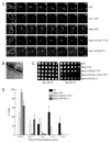
The synthetic lethality between cin8 and ipl1–315 mutants is not due to abrogation of the spindle checkpoint. (A) Five-fold serial dilutions of wild-type (SBY3), cin8Δ (SBY2198), and deg-cin8 (SBY3884) cells were plated at 30 and 37 °C on galactose media. (B) Wild-type (SBY3376), cin8Δ (SBY4652), and deg-cin8 (SBY3884) cells containing Spc42-GFP were released from G1 into non-permissive conditions at 30 °C. Samples were collected at the indicated time points, fixed and scored for budding (left graph) and the number of Spc42-GFP foci (right graph) (n=100). Two foci of Spc42-GFP were scored as separated SPBs. Note: The number of wild-type cells containing separated SPBs decreased over time because the cells entered G1 of the next cell cycle. (C) Five-fold serial dilutions of wild-type (SBY3), deg-cin8 (SBY3884), deg-cin8 ipl1–315 (SBY3887), deg-cin8 kip1Δ (SBY3964), and ipl1–315 kip1Δ (SBY5836) cells were plated at 30 °C on glucose and galactose media. (D) Wild-type (SBY818), deg-cin8 (SBY2642), deg-cin8 ipl1–321 (SBY2641), and deg-cin8 ipl1–315 (SBY2680) cells containing Pds1-myc were released from G1 at 30 °C into non-permissive conditions. Lysates were immunoblotted with anti-myc antibody.
Figure 4
Figure 4. Deg-cin8 ipl1–315
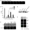
cells are defective in SPB separation. (A) Live microscopy was performed on wild-type (SBY3376), ipl1–315 (SBY5113), deg-cin8 (SBY3884), deg-cin8 ipl1–315 (SBY3887), and deg-cin8 kip1Δ (SBY3964) mutant cells expressing Spc42-GFP that were released to the non-permissive conditions from G1 at 30 °C. Time lapse microscopy was initiated 60 minutes after release and eight z-sections (0.4 μm intervals) were acquired every 40 s for 90 minutes. Images of single cells from the first 14 minutes of the time-lapse are shown (n=10). An outline of the cell is shown at 0 min. Scale bar equals 5 μm. (B) Transmission EM was performed on serial sections of deg-cin8 ipl1–315 cells that were released from G1 into non-permissive conditions for 90 minutes (n=14). A representative image is shown. (C) Five-fold serial dilutions of wild-type (SBY818), deg-cin8 (SBY 3884), deg-cin8 ipl1–315 (SBY3887), deg-cin8 ipl1–315 kip1Δ (SBY3970), and deg-cin8 kip1Δ (SBY3964) cells were plated at 26 °C on glucose and galactose media. (D) The distance between SPBs was measured for 10 cells from each of the strains from (A) every 2 minutes for a 20 minute time span. The measurements for each strain were compiled and the distributions based on pole-to-pole distance were plotted.
Figure 5
Figure 5
Ase1 is required for spindle assembly. (A) Live microscopy was performed on deg-cin8 ase1Δ mutant cells expressing Spc42-GFP (SBY4317) as described in Fig. 4A (n=10). (B) Microtubule morphology in wild-type (SBY1159), deg-cin8 ipl1–315 (SBY3887), deg-cin8 ase1Δ (SBY4317) cells released from a G1 arrest into the cell cycle for 150’ was analyzed by indirect immunofluorescence. (C) Five-fold serial dilutions of wild-type (SBY3), deg-cin8 (SBY3884), deg-cin8 ipl1–315 (SBY3887), and deg-cin8 ipl1–315 pGAL-ASE1 (SBY4382) cells were plated at 30 °C on glucose and galactose media. (D) ASE1 overexpression in deg-cin8 ipl1–315 cells rescues the SPB separation defect. Live microscopy was performed on deg-cin8 ipl1–315 pGAL-ASE1 (SBY4382) as described in Fig. 4A (n=10). Scale bar equals 5 μm on all panels.
Figure 6
Figure 6
Ase1 phosphorylation and localization depend on Ipl1. (A) and (B) Live microscopy was performed on deg-cin8 ase1Δ cells expressing Spc42-GFP that contain ase1–5A (SBY4903) 60 minutes after release from G1 at 30 °C. (B) The distance between SPBs was measured for 10 cells from each of the strains from (A) every 2 minutes for a 20 minute timespan. (C) Wild-type cells (SBY1159), ase1Δ cells (SBY5252), and ase1Δ cells with ASE1 (SBY5251) or ase1–5A (SBY5282) expressing Tub1-GFP were arrested in G1 and released to 30 °C. Microscopy was performed 90 min after release and a representative image of the phenotype of the majority of cells in each strain is shown (n=100). Scale bar equals 5 μm. (D) Wild-type (SBY1308), ipl1–321 (SBY2395), glc7–10 (SBY1305), glc7–10 ipl1–321 (SBY1994) cells were shifted to 37 °C for 2 h and Ase1 mobility was analyzed by immunoblotting. (E) Microscopy was performed on asynchronously growing wild-type (SBY5243) and ipl1–315 (SBY5242) cells expressing Ase1-GFP and Spc29-CFP. Small-budded cells with unseparated SPBs were scored for the presence (top panel) or absence (bottom panel) of Ase1 signal (n=100).
Figure 7
Figure 7
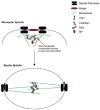
A model for Ipl1’s role in spindle assembly. Following SPB duplication, short MTs arising from the SPBs interdigitate over the bridge structure and are crosslinked by the MT bundling protein Ase1 and other MAPs. Ipl1 phosphorylation of Ase1 may increase Ase1’s affinity for MTs or could enhance Ase1’s specificity toward cross-linking anti-parallel MTs. Once MTs arising from the two SPBs are crosslinked by the midzone, motor proteins generate the outward forces that push the SPBs apart to generate a bipolar spindle and are balanced by the minus-end directed Kar3 kinesin.

Similar articles

Cited by

References

    1. Biggins S, Murray AW. The budding yeast protein kinase Ipl1/Aurora allows the absence of tension to activate the spindle checkpoint. Genes Dev. 2001;15:3118–3129. - PMC - PubMed
    1. Biggins S, Severin FF, Bhalla N, Sassoon I, Hyman AA, Murray AW. The conserved protein kinase Ipl1 regulates microtubule binding to kinetochores in budding yeast. Genes Dev. 1999;13:532–544. - PMC - PubMed
    1. Bishop AC, Ubersax JA, Petsch DT, Matheos DP, Gray NS, Blethrow J, Shimizu E, Tsien JZ, Schultz PG, Rose MD, et al. A chemical switch for inhibitor-sensitive alleles of any protein kinase. Nature. 2000;407:395–401. - PubMed
    1. Buvelot S, Tatsutani SY, Vermaak D, Biggins S. The budding yeast Ipl1/Aurora protein kinase regulates mitotic spindle disassembly. J Cell Biol. 2003;160:329–339. - PMC - PubMed
    1. Byers B, Goetsch L. Behavior of Spindle and Spindle Plaques in the Cell Cycle and Conjugation of Saccharomyces cerevisiae. J Bacteriol. 1975;124:511–523. - PMC - PubMed

Publication types

MeSH terms

Substances