Review
. 2007:163:63-84.
doi: 10.1016/S0079-6123(07)63004-0.

Extrinsic afferent systems to the dentate gyrus

Affiliations
Review

Extrinsic afferent systems to the dentate gyrus

Csaba Leranth et al. Prog Brain Res. 2007.

Abstract

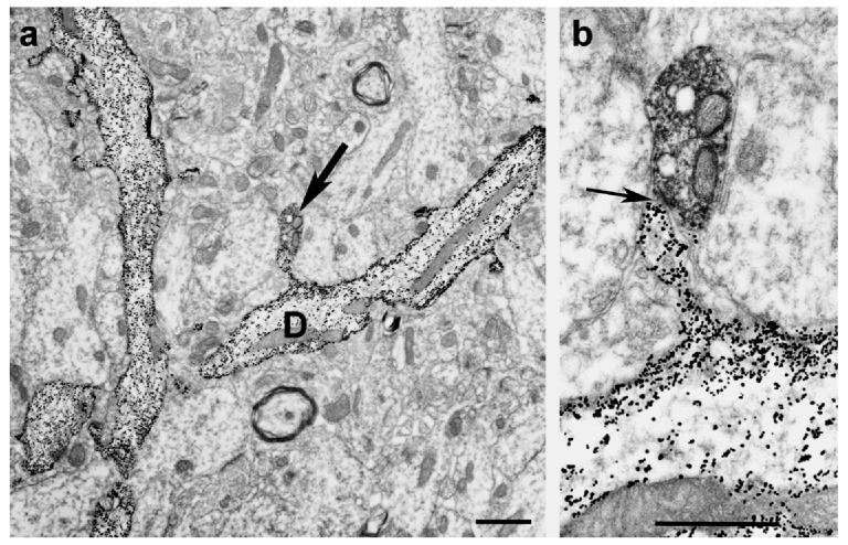
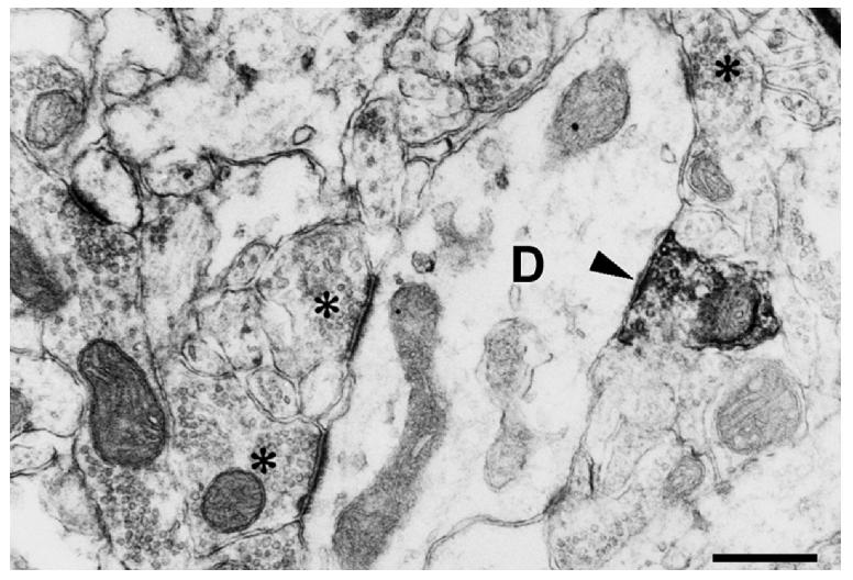
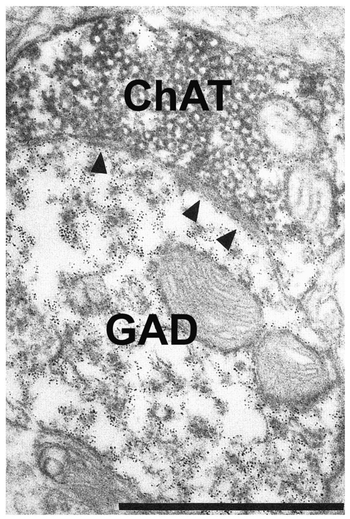
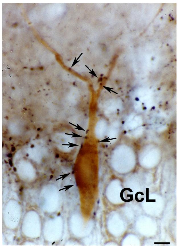
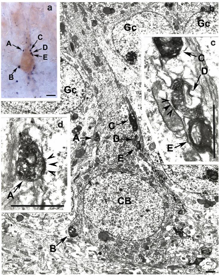
The dentate gyrus is the first stage of the intrahippocampal, excitatory, trisynaptic loop, and a primary target of the majority of entorhinal afferents that terminate in a laminar fashion on granule cell dendrites and carry sensory information of multiple modalities about the external world. The electric activity of the trisynaptic pathway is controlled mainly by different types of local, GABAergic interneurons, and subcortical and commissural afferents. In this chapter we will outline the origin and postsynaptic targets in the dentate gyrus of chemically identified subcortical inputs. These systems are afferents originating from the medial septum/diagonal band of Broca GABAergic and cholinergic neurons, neurochemically distinct types of neurons located in the supramammillary area, serotonergic fibers from the median raphe, noradrenergic afferents from the pontine nucleus, locus ceruleus, dopamine axons originating in the ventral tegmental area, and the commissural projection system. Because of the physiological implications, these afferents are discussed in the context of the glutamatergic innervation of the dentate gyrus. One common feature of the extrinsic dentate afferent systems is that they originate from a relatively small number of neurons. However, the majority of these afferents are able to exert a powerful control over the electrical activity of the hippocampus. This strong influence is due to the fact that the majority of the extrinsic afferents terminate on a relatively small, but specific, populations of neurons that are able to control large areas of the hippocampal formation.


Figures

Fig. 1
Fig. 1
Low (Panel a) and high power (Panel b) electron micrographs (received from Dr. Michael Frotscher) show the result of a combined Golgi impregnation and ChAT immunostaining experiment. On Panel a, Golgi-impregnated (gold-toned) spiny granule cell dendrites are seen. Arrow on the same panel points at a ChAT immunoreactive bouton contacting the spine head of the dendrite (D). Panel b shows the asymmetric synaptic contact (arrow) between the two profiles. Bar scales= 1 μm.
Fig. 2
Fig. 2
Electron micrograph taken from the molecular layer of the dentate gyrus immunostained for ChAT. A ChAT-immunoreactive axon terminal forms asymmetric synaptic contact with a non-spiny dendritic shaft (D). The lack of dendritic spines indicate that this dendrite is the process of an interneuron. Asterisks label axon terminals forming asymmetric synapses with the same dendrite. Bar scale= 1 μm.
Fig. 3
Fig. 3
Light micrograph (kindly provided by Dr. Attila Gulyas) shows the result of a combined anterograde tracing and calretinin immunostaining study, in the dentate gyrus. The anterograde tracer, biotinylated dextran amine (BDA) was injected into the medial septum diagonal band. Large boutons of BDA-containing axons form multiple, basket-like, putative synaptic contacts with the soma of calretinin-immunoreactive (labeled with a brown diaminobenzidine reaction product) neurons. One of these cells (arrows) located in the supragranular layer (SgL) the other is at the border between the granule cell layer (GcL) and dentate hilar area (H). Bar scale= 50 μm.
Fig. 4
Fig. 4
Electron micrograph shows the result of a double immunostaining experiment for choline acetyltransferase (ChAT) and glutamic acid decarboxylase (GAD), in the rat medial septum diagonal band of Broca. Immunoreactivity for ChAT and GAD was visualized with two contrasting immunomarker, diaminobenzidine reaction and ferritin labeling, respectively. A ChAT-immunoreactive bouton form asymmetric synaptic contact (arrowheads) with a GAD immunoreactive dendrite. Bar scale= 1 μm.
Fig. 5
Fig. 5
Light micrograph taken from a double immunostained vibratome section of the monkey dentate gyrus. Immunoreactivity for substance P was labeled with a dark-blue Ni-diaminobenzidine reaction, while immunostaining for parvalbumin was visualized by a brown diaminobenzidine reaction. The soma and dendrites of the parvalbumin-containing cell embedded into the granule cell layer (GcL) is contacted by several substance P-immunoreactive axon terminals (arrows). Bar scale= 10 μm.
Fig. 6
Fig. 6
Light (Panel a) and electron micrographs (Panels b, c, d) depicted from the monkey dentate gyrus demonstrate the result of a correlated light and electron microscopic double immunostaining for substance P (labeled by a dark-blue blue Ni-diaminobenzidine reaction) and calbindin (brown diaminobenzidine chromogen). Panel a shows a calbindin-immunoreactive neuron embedded into the granule cell layer forming putative synaptic contacts (A – D) with substance P-containing axon terminals. Electron microscopic analysis of ultrathin sections cut from the same area shows that boutons A and D form robust asymmetric synaptic contacts (arrowheads on panels c and d) with the soma of this calbindin-containing cell (CB on panel b). Gc- granule cell. Bar scales= panel a, 10 μm; panels b – c, 1 μm.

References

    1. Aihara Y, Mashima H, Onda H, Hisano S, Kasuya H, Hori T, Yamada S, Tomura H, Yamada Y, Inoue I, Kojima I, Takeda J. Molecular cloning of a novel brain-type Na(+)-dependent inorganic phosphate cotransporter. J. Neurochem. 2000;74(6):2622–2625. - PubMed
    1. Alreja M. Excitatory actions of serotonin on GABAergic neurons of the medial septum diagonal band of Broca. Synapse. 1966;22:15–27. - PubMed
    1. Amaral DG. A Golgi study of cell types in the hilar region of the hippocampus in the rat. J Comp Neurol. 1978;182:851–914. - PubMed
    1. Amaral DG, Cowan WM. Subcortical afferents to the hippocampal formation in the monkey. J. Camp. Neurol. 1980;189:573–591. - PubMed
    1. Amaral DG, Witter MP. The three-dimensional organization of the hippocampal formation: a review of anatomical data. Neuroscience. 1989;31:571–591. - PubMed

Publication types

MeSH terms

LinkOut - more resources