

Sex steroids and the dentate gyrus
- PMID: 17765731
- PMCID: PMC1964752
- DOI: 10.1016/S0079-6123(07)63023-4
Sex steroids and the dentate gyrus
Abstract
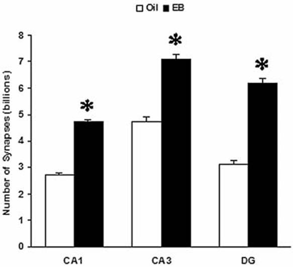
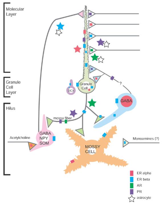
In the late 1980s, the finding that the dentate gyrus contains more granule cells in the male than in the female of certain mouse strains provided the first indication that the dentate gyrus is a significant target for the effects of sex steroids during development. Gonadal hormones also play a crucial role in shaping the function and morphology of the adult brain. Besides reproduction-related processes, sex steroids participate in higher brain operations such as cognition and mood, in which the hippocampus is a critical mediator. Being part of the hippocampal formation, the dentate gyrus is naturally involved in these mechanisms and as such, this structure is also a critical target for the activational effects of sex steroids. These activational effects are the results of three major types of steroid-mediated actions. Sex steroids modulate the function of dentate neurons under normal conditions. In addition, recent research suggests that hormone-induced cellular plasticity may play a larger role than previously thought, particularly in the dentate gyrus. Specifically, the regulation of dentate gyrus neurogenesis and synaptic remodeling by sex steroids received increasing attention lately. Finally, the dentate gyrus is influenced by gonadal hormones in the context of cellular injury, and the work in this area demonstrates that gonadal hormones have neuroprotective potential. The expression of estrogen, progestin, and androgen receptors in the dentate gyrus suggests that sex steroids, which could be of gonadal origin and/or synthesized locally in the dentate gyrus, may act directly on dentate cells. In addition, gonadal hormones could also influence the dentate gyrus indirectly, by subcortical hormone-sensitive structures such as the cholinergic septohippocampal system. Importantly, these three sex steroid-related themes, functional effects in the normal dentate gyrus, mechanisms involving neurogenesis and synaptic remodeling, as well as neuroprotection, have substantial implications for understanding normal cognitive function, with clinical importance for epilepsy, Alzheimer's disease and mental disorders.
Figures
Similar articles
-
Gonadal hormone modulation of neurogenesis in the dentate gyrus of adult male and female rodents.Brain Res Rev. 2008 Mar;57(2):332-41. doi: 10.1016/j.brainresrev.2007.05.008. Epub 2007 Jun 9. Brain Res Rev. 2008. PMID: 17669502
-
Seasonal and sex differences in cell proliferation, neurogenesis, and cell death within the dentate gyrus of adult wild-caught meadow voles.Neuroscience. 2017 Sep 30;360:155-165. doi: 10.1016/j.neuroscience.2017.07.046. Epub 2017 Jul 28. Neuroscience. 2017. PMID: 28757249
-
Sex differences in the hippocampal dentate gyrus of the guinea-pig before puberty.Neuroscience. 2003;121(2):327-39. doi: 10.1016/s0306-4522(03)00434-2. Neuroscience. 2003. PMID: 14521992
-
Gonadal hormone modulation of hippocampal neurogenesis in the adult.Hippocampus. 2006;16(3):225-32. doi: 10.1002/hipo.20154. Hippocampus. 2006. PMID: 16411182 Review.
-
Gonadal and adrenal steroids regulate neurochemical and structural plasticity of the hippocampus via cellular mechanisms involving NMDA receptors.Cell Mol Neurobiol. 1996 Apr;16(2):103-16. doi: 10.1007/BF02088170. Cell Mol Neurobiol. 1996. PMID: 8743963 Free PMC article. Review.
Cited by
-
Apolipoprotein E4 causes age- and sex-dependent impairments of hilar GABAergic interneurons and learning and memory deficits in mice.PLoS One. 2012;7(12):e53569. doi: 10.1371/journal.pone.0053569. Epub 2012 Dec 31. PLoS One. 2012. PMID: 23300939 Free PMC article.
-
Nicotine Significantly Improves Chronic Stress-Induced Impairments of Cognition and Synaptic Plasticity in Mice.Mol Neurobiol. 2017 Aug;54(6):4644-4658. doi: 10.1007/s12035-016-0012-2. Epub 2016 Jul 12. Mol Neurobiol. 2017. PMID: 27405470
-
Hormonal mechanisms of cooperative behaviour.Philos Trans R Soc Lond B Biol Sci. 2010 Sep 12;365(1553):2737-50. doi: 10.1098/rstb.2010.0151. Philos Trans R Soc Lond B Biol Sci. 2010. PMID: 20679116 Free PMC article. Review.
-
Stress experienced in utero reduces sexual dichotomies in neurogenesis, microenvironment, and cell death in the adult rat hippocampus.Dev Neurobiol. 2008 Apr;68(5):575-89. doi: 10.1002/dneu.20600. Dev Neurobiol. 2008. PMID: 18264994 Free PMC article.
-
The Enigma of the Adrenarche: Identifying the Early Life Mechanisms and Possible Role in Postnatal Brain Development.Int J Mol Sci. 2021 Apr 21;22(9):4296. doi: 10.3390/ijms22094296. Int J Mol Sci. 2021. PMID: 33919014 Free PMC article. Review.
References
-
- Altman J, Das GD. Autoradiographic and histological evidence of postnatal hippocampal neurogenesis in rats. J. Comp. Neurol. 1965;124(3):319–335. - PubMed
-
- Azcoitia I, Sierra A, Garcia-Segura LM. Estradiol prevents kainic acid-induced neuronal loss in the rat dentate gyrus. Neuroreport. 1998;9(13):3075–3079. - PubMed
-
- Azcoitia I, Sierra A, Garcia-Segura LM. Neuroprotective effects of estradiol in the adult rat hippocampus: interaction with insulin-like growth factor-I signalling. J. Neurosci. Res. 1999a;58(6):815–822. - PubMed
-
- Azcoitia I, Sierra A, Garcia-Segura LM. Localization of estrogen receptor beta-immunoreactivity in astrocytes of the adult rat brain. Glia. 1999b;26(3):260–267. - PubMed
-
- Banasr M, Hery M, Brezun JM, Daszuta A. Serotonin mediates oestrogen stimulation of cell proliferation in the adult dentate gyrus. Eur. J. Neurosci. 2001;14(9):1417–1424. - PubMed
Publication types
MeSH terms
Substances
Grants and funding
LinkOut - more resources
Full Text Sources
