

Visuomotor contribution to force variability in the plantarflexor and dorsiflexor muscles
- PMID: 17765988
- PMCID: PMC2148254
- DOI: 10.1016/j.humov.2007.07.001
Visuomotor contribution to force variability in the plantarflexor and dorsiflexor muscles
Abstract
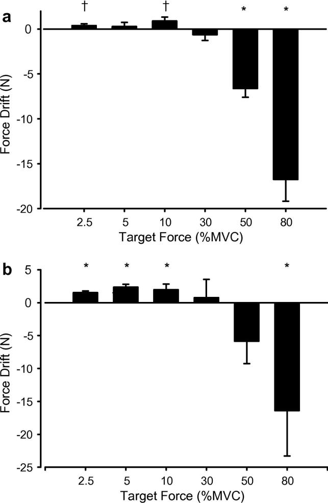
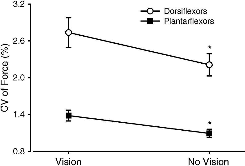
The visual correction employed during isometric contractions of large proximal muscles contributes variability to the descending command and alters fluctuations in muscle force. This study explored the contribution of visuomotor correction to isometric force fluctuations for the more distal dorsiflexor (DF) and plantarflexor (PF) muscles of the ankle. Twenty-one healthy adults performed steady isometric contractions with the DF and PF muscles both with (VIS) and without (NOVIS) visual feedback of the force. The target forces exerted ranged from 2.5% to 80% MVC. The standard deviation (SD) and coefficient of variation (CV) of force was measured from the detrended (drift removed) VIS and NOVIS steadiness trials. Removal of VIS reduced the CV of force by 19% overall. The reduction in fluctuations without VIS was significant across a large range of target forces and was more consistent for the PF than the DF muscles. Thus, visuomotor correction contributes to the variability of force during isometric contractions of the ankle dorsiflexors and plantarflexors.
Figures


Similar articles
-
Force control is impaired in the ankle plantarflexors of elderly adults.Eur J Appl Physiol. 2007 Nov;101(5):629-36. doi: 10.1007/s00421-007-0538-0. Epub 2007 Aug 15. Eur J Appl Physiol. 2007. PMID: 17701201
-
Variability of quadriceps femoris motor neuron discharge and muscle force in human aging.Exp Brain Res. 2007 May;179(2):219-33. doi: 10.1007/s00221-006-0785-z. Epub 2006 Nov 30. Exp Brain Res. 2007. PMID: 17136528
-
Visuomotor Correction is a Robust Contributor to Force Variability During Index Finger Abduction by Older Adults.Front Aging Neurosci. 2015 Dec 15;7:229. doi: 10.3389/fnagi.2015.00229. eCollection 2015. Front Aging Neurosci. 2015. PMID: 26696881 Free PMC article.
-
Aging, visuomotor correction, and force fluctuations in large muscles.Med Sci Sports Exerc. 2007 Mar;39(3):469-79. doi: 10.1249/mss.0b013e31802d3ad3. Med Sci Sports Exerc. 2007. PMID: 17473773
-
The length of tibialis anterior does not influence force steadiness during submaximal isometric contractions with the dorsiflexors.Eur J Sport Sci. 2022 Apr;22(4):539-548. doi: 10.1080/17461391.2021.1922506. Epub 2021 May 21. Eur J Sport Sci. 2022. PMID: 33899692
Cited by
-
Does pain influence force steadiness? A protocol for a systematic review.BMJ Open. 2021 Jan 8;11(1):e042525. doi: 10.1136/bmjopen-2020-042525. BMJ Open. 2021. PMID: 33419915 Free PMC article.
-
Removing visual feedback for a single limb alters between-limb force tremor relationships during isometric bilateral contractions.Exp Brain Res. 2015 Jan;233(1):115-24. doi: 10.1007/s00221-014-4098-3. Epub 2014 Sep 19. Exp Brain Res. 2015. PMID: 25234402
-
Editorial: A Hand at Work: Effects of Aging.Front Aging Neurosci. 2016 Jun 14;8:141. doi: 10.3389/fnagi.2016.00141. eCollection 2016. Front Aging Neurosci. 2016. PMID: 27378913 Free PMC article. No abstract available.
-
Force variability is mostly not motor noise: Theoretical implications for motor control.PLoS Comput Biol. 2021 Mar 8;17(3):e1008707. doi: 10.1371/journal.pcbi.1008707. eCollection 2021 Mar. PLoS Comput Biol. 2021. PMID: 33684099 Free PMC article.
-
Visual feedback improves bimanual force control performances at planning and execution levels.Sci Rep. 2021 Oct 27;11(1):21149. doi: 10.1038/s41598-021-00721-9. Sci Rep. 2021. PMID: 34707163 Free PMC article.
References
-
- Bawa P, Chalmers GR, Stewart H, Eisen AA. Responses of ankle extensor and flexor motoneurons to transcranial magnetic stimulation. Journal of Neurophysiology. 2002;88:124–132. - PubMed
-
- Belanger AY, McComas AJ, Elder GB. Physiological properties of two antagonist human muscle groups. European Journal of Applied Physiology and Occupational Physiology. 1983;51:381–393. - PubMed
-
- Brouwer B, Ashby P. Corticospinal projections to upper and lower limb spinal motoneurons in man. Electroencephalography and Clinical Neurophysiology. 1990;76:509–519. - PubMed
-
- Calvin WH, Stevens CF. Synaptic noise and other sources of randomness in motoneuron interspike intervals. Journal of Neurophysiology. 1968;31:574–587. - PubMed
-
- Capaday C, Lavoie BA, Barbeau H, Schneider C, Bonnard M. Studies on the corticospinal control of human walking. I. Responses to focal transcranial magnetic stimulation of the motor cortex. Journal of Neurophysiology. 1999;81:129–139. - PubMed
Publication types
MeSH terms
Grants and funding
LinkOut - more resources
Full Text Sources
Miscellaneous
