

On navigating the human cerebral cortex: response to 'in praise of tedious anatomy'
- PMID: 17766148
- PMCID: PMC2045137
- DOI: 10.1016/j.neuroimage.2007.02.021
On navigating the human cerebral cortex: response to 'in praise of tedious anatomy'
Abstract
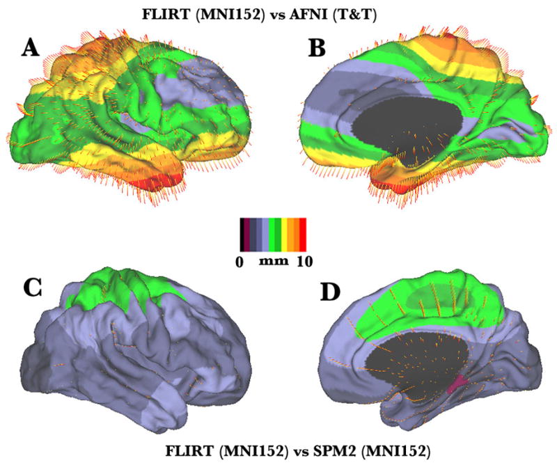
Individual variability of the human cerebral cortex is a source of both fascination and frustration. The fascination arises because variability in cortical structure and function may account for many aspects of our unique personalities and cognitive capabilities. For neuroimagers, the frustration arises because variability presents serious obstacles when attempting to assign particular functional activation patterns to specific cortical areas. Devlin and Poldrack cogently summarize many of the key issues, and they make useful suggestions for linking function to anatomy using a standardized stereotaxic space. This commentary provides a broader perspective on the nature of individual variability that has implications for the choice of strategies used to compensate for variability. It also includes information about the actual differences between various registration strategies and introduces a new strategy for converting neuroimaging data to a standard stereotaxic space.
Figures
Comment on
-
In praise of tedious anatomy.Neuroimage. 2007 Oct 1;37(4):1033-41; discussion 1050-8. doi: 10.1016/j.neuroimage.2006.09.055. Neuroimage. 2007. PMID: 17870621 Free PMC article. Review.
Similar articles
-
Accurate prediction of V1 location from cortical folds in a surface coordinate system.Neuroimage. 2008 Feb 15;39(4):1585-99. doi: 10.1016/j.neuroimage.2007.10.033. Epub 2007 Nov 6. Neuroimage. 2008. PMID: 18055222 Free PMC article.
-
Automatic 3D intersubject registration of MR volumetric data in standardized Talairach space.J Comput Assist Tomogr. 1994 Mar-Apr;18(2):192-205. J Comput Assist Tomogr. 1994. PMID: 8126267
-
Detection and mapping of abnormal brain structure with a probabilistic atlas of cortical surfaces.J Comput Assist Tomogr. 1997 Jul-Aug;21(4):567-81. doi: 10.1097/00004728-199707000-00008. J Comput Assist Tomogr. 1997. PMID: 9216760
-
Automated extraction and variability analysis of sulcal neuroanatomy.IEEE Trans Med Imaging. 1999 Mar;18(3):206-17. doi: 10.1109/42.764891. IEEE Trans Med Imaging. 1999. PMID: 10363699 Review.
-
Advances in cytoarchitectonic mapping of the human cerebral cortex.Neuroimaging Clin N Am. 2001 May;11(2):151-69, vii. Neuroimaging Clin N Am. 2001. PMID: 11489732 Review.
Cited by
-
Structure-function relationship of working memory activity with hippocampal and prefrontal cortex volumes.Brain Struct Funct. 2013 Jan;218(1):173-86. doi: 10.1007/s00429-012-0391-8. Epub 2012 Feb 24. Brain Struct Funct. 2013. PMID: 22362200 Free PMC article.
-
Functional Parcellation of the Cerebral Cortex Across the Human Adult Lifespan.Cereb Cortex. 2018 Dec 1;28(12):4403-4423. doi: 10.1093/cercor/bhy218. Cereb Cortex. 2018. PMID: 30307480 Free PMC article.
-
The prosubiculum in the human hippocampus: A rostrocaudal, feature-driven, and systematic approach.J Comp Neurol. 2024 Mar;532(3):e25604. doi: 10.1002/cne.25604. J Comp Neurol. 2024. PMID: 38477395 Free PMC article.
-
Symmetric abnormalities in sulcal patterning in schizophrenia.Neuroimage. 2008 Nov 15;43(3):440-6. doi: 10.1016/j.neuroimage.2008.07.034. Epub 2008 Jul 29. Neuroimage. 2008. PMID: 18707008 Free PMC article.
-
Using transcranial direct-current stimulation (tDCS) to understand cognitive processing.Atten Percept Psychophys. 2017 Jan;79(1):3-23. doi: 10.3758/s13414-016-1224-2. Atten Percept Psychophys. 2017. PMID: 27804033 Free PMC article. Review.
References
-
- Amunts K, Schleicher A, Burgel U, Mohlberg H, Uylings HB, Zilles K. Broca’s region revisited: Cytoarchitecture and intersubject variability. J Comp Neurol. 1999;412:319–341. - PubMed
-
- Amunts K, Malikovic A, Mohlberg H, Schormann T, Zilles K. Brodmann’s areas 17 and 18 brought into stereotaxic space -- Where and how variable? NeuroImage. 2000;11:66–84. - PubMed
-
- Amunts K, Weiss PH, Mohlberg H, Pieperhoff P, Eickhoff S, Gurd JM, Marshall JC, Shah NJ, Fing GR, Zilles K. Analysis of neural mechanisms underlying verbal fluency in cytoarchitectonically defined stereotaxic space – The roles of Brodmann areas 44 and 45. NeuroImage. 2004;22:42–56. - PubMed
Publication types
MeSH terms
Grants and funding
LinkOut - more resources
Full Text Sources
