

Singlet oxygen photosensitization by EGFP and its chromophore HBDI
- PMID: 17766345
- PMCID: PMC2134865
- DOI: 10.1529/biophysj.107.107128
Singlet oxygen photosensitization by EGFP and its chromophore HBDI
Abstract
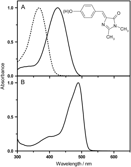
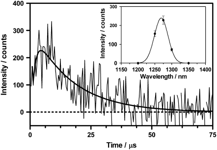
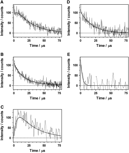
The photosensitization of reactive oxygen species and, in particular, singlet oxygen by proteins from the green fluorescent protein (GFP) family influences important processes such as photobleaching and genetically targeted chromophore-assisted light inactivation. In this article, we report an investigation of singlet oxygen photoproduction by GFPs using time-resolved detection of the NIR phosphorescence of singlet oxygen at 1275 nm. We have detected singlet oxygen generated by enhanced (E)GFP, and measured a lifetime of 4 micros in deuterated solution. By comparison with the model compound of the EGFP fluorophore 4-hydroxybenzylidene-1,2-dimethylimidazoline (HBDI), our results confirm that the beta-can of EGFP provides shielding of the fluorophore and reduces the production of this reactive oxygen species. In addition, our results yield new information about the triplet state of these proteins. The quantum yield for singlet oxygen photosensitization by the model chromophore HBDI is 0.004.
Figures
Similar articles
-
Singlet oxygen photosensitisation by GFP mutants: oxygen accessibility to the chromophore.Photochem Photobiol Sci. 2010 Oct 28;9(10):1336-41. doi: 10.1039/c0pp00125b. Epub 2010 Jul 30. Photochem Photobiol Sci. 2010. PMID: 20672172
-
Chromophore-assisted laser inactivation in cell biology.Trends Cell Biol. 2008 Sep;18(9):443-50. doi: 10.1016/j.tcb.2008.07.001. Epub 2008 Aug 14. Trends Cell Biol. 2008. PMID: 18706812 Free PMC article. Review.
-
Singlet oxygen photosensitisation by the fluorescent probe Singlet Oxygen Sensor Green.Chem Commun (Camb). 2009 May 28;(20):2920-2. doi: 10.1039/b822776d. Epub 2009 Apr 1. Chem Commun (Camb). 2009. PMID: 19436910
-
Effect of sensitizer protonation on singlet oxygen production in aqueous and nonaqueous media.J Phys Chem A. 2007 May 31;111(21):4573-83. doi: 10.1021/jp066843f. Epub 2007 May 5. J Phys Chem A. 2007. PMID: 17480060
-
Riboflavin as a photosensitizer. Effects on human health and food quality.Food Funct. 2012 May;3(5):487-502. doi: 10.1039/c2fo10246c. Epub 2012 Mar 12. Food Funct. 2012. PMID: 22406738 Review.
Cited by
-
Understanding the Dynamics of the Transient and Permanent Opening Events of the Mitochondrial Permeability Transition Pore with a Novel Stochastic Model.Membranes (Basel). 2022 Apr 30;12(5):494. doi: 10.3390/membranes12050494. Membranes (Basel). 2022. PMID: 35629820 Free PMC article.
-
Autofluorescent proteins as photosensitizer in eukaryontes.Int J Med Sci. 2009 Dec 1;6(6):365-73. doi: 10.7150/ijms.6.365. Int J Med Sci. 2009. PMID: 19960122 Free PMC article.
-
Positioning effects of KillerRed inside of cells correlate with DNA strand breaks after activation with visible light.Int J Med Sci. 2011 Jan 21;8(2):97-105. doi: 10.7150/ijms.8.97. Int J Med Sci. 2011. PMID: 21278894 Free PMC article.
-
Self-labelling enzymes as universal tags for fluorescence microscopy, super-resolution microscopy and electron microscopy.Sci Rep. 2015 Dec 8;5:17740. doi: 10.1038/srep17740. Sci Rep. 2015. PMID: 26643905 Free PMC article.
-
Direct visualization of protein association in living cells with complex-edited electron microscopy.Angew Chem Int Ed Engl. 2010 Oct 18;49(43):7952-4. doi: 10.1002/anie.201003217. Angew Chem Int Ed Engl. 2010. PMID: 20845344 Free PMC article. No abstract available.
References
-
- Giepmans, B. N. G., S. R. Adams, M. H. Ellisman, and R. Y. Tsien. 2006. The fluorescent toolbox for assessing protein location and function. Science. 312:217–224. - PubMed
-
- Remington, S. J. 2006. Fluorescent proteins: maturation, photochemistry and photophysics. Curr. Opin. Struct. Biol. 16:714–721. - PubMed
-
- Dixit, R., and R. Cyr. 2003. Cell damage and reactive oxygen species production induced by fluorescence microscopy: effect on mitosis and guidelines for non-invasive fluorescence microscopy. Plant J. 36:280–290. - PubMed
-
- Greenbaum, L., C. Rothmann, R. Lavie, and Z. Malik. 2000. Green fluorescent protein photobleaching: a model for protein damage by endogenous and exogenous singlet oxygen. Biol. Chem. 381:1251–1258. - PubMed
-
- Halliwell, B., and J. M. C. Gutteridge. 1999. Free Radicals in Biology and Medicine. Oxford University Press, Oxford, UK.
Publication types
MeSH terms
Substances
LinkOut - more resources
Full Text Sources
Other Literature Sources
Miscellaneous
