. 2007 Nov;189(21):7896-910.
doi: 10.1128/JB.00726-07. Epub 2007 Aug 31.

Antigen 84, an effector of pleiomorphism in Mycobacterium smegmatis

Affiliations

Antigen 84, an effector of pleiomorphism in Mycobacterium smegmatis

Liem Nguyen et al. J Bacteriol. 2007 Nov.

Abstract

While in most rod-shaped bacteria, morphology is based on MreB-like proteins that form an actin-like cytoskeletal scaffold for cell wall biosynthesis, the factors that determine the more flexible rod-like shape in actinobacteria such as Mycobacterium species are unknown. Here we show that a Mycobacterium smegmatis protein homologous to eubacterial DivIVA-like proteins, including M. tuberculosis antigen 84 (Ag84), localized symmetrically to centers of peptidoglycan biosynthesis at the poles and septa. Controlled gene disruption experiments indicated that the gene encoding Ag84, wag31, was essential; when overexpressed, cells became longer and wider, with Ag84 asymmetrically distributed at one pole. Many became grossly enlarged, bowling-pin-shaped cells having up to 80-fold-increased volume. In these cells, Ag84 accumulated predominantly at a bulbous pole that was apparently generated by uncontrolled cell wall expansion. In some cells, Ag84 was associated with exceptional sites of cell wall expansion (buds) that evolved into branches. M. bovis BCG Ag84 was able to form oligomers in vitro, perhaps reflecting its superstructure in vivo. These data suggested a role for Ag84 in cell division and modulating cell shape in pleiomorphic actinobacteria.


Figures
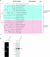
FIG. 1.
FIG. 1.
The DivIVA homolog in mycobacteria. (A) Phylogenetic representation of DivIVA-like proteins from gram-positive bacteria in relation to morphology and the presence of MreB-like proteins. In firmicutes, the presence of MreB correlates with a rod shape, while in most rod-like, sometimes pleiomorphic, actinobacteria, MreB is not present. Protein sequences were obtained from the National Center for Biotechnology Information (http://www.ncbi.nlm.nih.gov/genomes/lproks.cgi) and the TIGR genome databases (www.tigr.org/). Alignment of protein sequences was carried out using the ClustalW algorithm (62). The tree was generated using the UPGMA algorithm in the MacVector 8.0 package (Accelrys Inc., San Diego, CA). A distance bar is shown under the tree. (B) The M. smegmatis gene encoding Ag84MS (wag31MS) is essential. The chromosomal wag31MS could only be disrupted by phage-mediated allelic exchange when wild-type M. smegmatis was supplemented in trans with a vector expressing wag31MS from the acetamidase promoter. The disruption was confirmed by recovering the mutant locus by PCR using primers flanking the putative open reading frame. The mutant gene containing the inserted hygromycin resistance cassette generated a larger fragment (left panel). Ag84MS expression of the chromosomal wag31MS-deleted mutant was detected by Western blotting using antibody F126-2 against Ag84. Expression of Ag84MS from the acetamidase promoter without addition of the inducer acetamide was higher than wild-type expression levels (right panel). The sizes of molecular weight markers used for Western blots are indicated.
FIG. 2.
FIG. 2.
Ag84MS is recruited to polar sites of cell wall extension during growth and to midcell during septation. Wild-type M. smegmatis cells were transformed with pNDL2, generating a mutation in which wag31MS-GFP (encoding the Ag84MS-GFP fusion protein) was expressed from its native genomic promoter. Transformants grown on selective plates for 4 days were prepared for microscopic analyses. (A) Localization of Ag84. M. smegmatis cells expressing Ag84MS-GFP (pNDL2) were photographed under a fluorescence microscope (upper panels). In the majority of cells, fluorescence was observed at the cell poles. In elongated cells, Ag84MS-GFP was also detected at the midcell. Bar, 1 μm. Polar localization was also observed by TEM using a monoclonal antibody against Ag84 (lower panels) to visualize Ag84 in cross-sections of cells. Arrowheads indicate positions of gold particles. Bars in lower panels, 0.5 μm. (B) Time-lapse video microscopy of Mycobacterium smegmatis mc2155 expressing wag31MS-gfp over a period of 180 min. A representative series of images of a cell size extension event (cell 1) and a cell division event (cell 2) viewed with differential interference contrast (DIC) (upper panels) and fluorescence (lower panels) microscopy are shown. Sites of extension and division are indicated by filled and empty arrows, respectively. Time points are indicated in minutes. Cartoon interpretations are shown below the panels. (C) Nascent peptidoglycan synthesis is localized at the poles, as revealed by staining growing cell walls with fluorescent vancomycin. M. smegmatis wild-type cells cultured on 7H10 agar medium for 4 days at 37°C were labeled and observed by use of a fluorescence microscope as described in Materials and Methods.
FIG. 3.
FIG. 3.
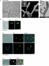
Overexpression of wag31MS altered the morphology of M. smegmatis cells. Wild-type M. smegmatis cells were transformed with pNDL4 overexpressing wag31MS or with pNDL1 in which the wag31MS-gfp (encoding the Ag84MS-GFP fusion protein) was overexpressed from Phsp60. (A) Enlargement and branching of M. smegmatis overexpressing wag31MS (pNDL4). After 2 days of growth on selective medium, transformants were harvested and observed. Left, middle, and right panels show phase-contrast, scanning electron microscopy (SEM), and TEM images of M. smegmatis overexpressing wag31MS. Insets: left panel, wild-type M. smegmatis mc2155 as control (bar in the inset, 1 μm); middle panel, relative shape and size of wild-type M. smegmatis mc2155 (bar in the inset, 0.5 μm); right panel, wild-type M. smegmatis mc2155 as control (bar in the inset, 0.91 μm). (B) Accumulations of Ag84MS-GFP (pNDL1) along the cylindrical part of cells were often associated with bud initiation (white arrows, top panels). Later, structures resembling emerging buds contained localized patches of Ag84MS (white arrows, middle panels). Emerging branches contained Ag84MS at the growing tip (white arrows, bottom panels). Bars, 3 μm. (C) Distorted heterogeneous shapes of M. smegmatis overexpressing wag31MS-gfp (pNDL1). After 3 days of growth on selective medium, transformants were viewed using differential interference contrast (DIC) and fluorescence modes. Similar results were obtained with cells overexpressing wag31MS alone. Minicells (arrows) and cells having rod, ovoid, round, or bottle-like shapes were observed. DAPI (4′,6′-diamidino-2-phenylindole) staining experiments showed that the majority of these small cells did not have DNA (data not shown). Bars, 10 μm. (D) By day 5 after transformation, M. smegmatis cells transformed with the wag31MS-gfp-overexpressing plasmid exhibited normal size and morphology. Bar, 2 μm. (E) Expression of Ag84MS in cell extracts of M. smegmatis strains cultured on 7H10 agar plates for 3 days at 37°C was detected by Western blot analysis with antibody against antigen 84 (F126-2; left panel) or GFP (right panel). The middle band in mc2155/Phsp60-wag31MS-gfp most likely represents a GFP degradation product of Ag84MS-GFP. The sizes of molecular weight markers used for Western blots are indicated.
FIG. 3.
FIG. 3.
Overexpression of wag31MS altered the morphology of M. smegmatis cells. Wild-type M. smegmatis cells were transformed with pNDL4 overexpressing wag31MS or with pNDL1 in which the wag31MS-gfp (encoding the Ag84MS-GFP fusion protein) was overexpressed from Phsp60. (A) Enlargement and branching of M. smegmatis overexpressing wag31MS (pNDL4). After 2 days of growth on selective medium, transformants were harvested and observed. Left, middle, and right panels show phase-contrast, scanning electron microscopy (SEM), and TEM images of M. smegmatis overexpressing wag31MS. Insets: left panel, wild-type M. smegmatis mc2155 as control (bar in the inset, 1 μm); middle panel, relative shape and size of wild-type M. smegmatis mc2155 (bar in the inset, 0.5 μm); right panel, wild-type M. smegmatis mc2155 as control (bar in the inset, 0.91 μm). (B) Accumulations of Ag84MS-GFP (pNDL1) along the cylindrical part of cells were often associated with bud initiation (white arrows, top panels). Later, structures resembling emerging buds contained localized patches of Ag84MS (white arrows, middle panels). Emerging branches contained Ag84MS at the growing tip (white arrows, bottom panels). Bars, 3 μm. (C) Distorted heterogeneous shapes of M. smegmatis overexpressing wag31MS-gfp (pNDL1). After 3 days of growth on selective medium, transformants were viewed using differential interference contrast (DIC) and fluorescence modes. Similar results were obtained with cells overexpressing wag31MS alone. Minicells (arrows) and cells having rod, ovoid, round, or bottle-like shapes were observed. DAPI (4′,6′-diamidino-2-phenylindole) staining experiments showed that the majority of these small cells did not have DNA (data not shown). Bars, 10 μm. (D) By day 5 after transformation, M. smegmatis cells transformed with the wag31MS-gfp-overexpressing plasmid exhibited normal size and morphology. Bar, 2 μm. (E) Expression of Ag84MS in cell extracts of M. smegmatis strains cultured on 7H10 agar plates for 3 days at 37°C was detected by Western blot analysis with antibody against antigen 84 (F126-2; left panel) or GFP (right panel). The middle band in mc2155/Phsp60-wag31MS-gfp most likely represents a GFP degradation product of Ag84MS-GFP. The sizes of molecular weight markers used for Western blots are indicated.
FIG. 4.
FIG. 4.
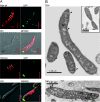
Ag84MS overexpression inhibited septum formation and created giant cells. (A) Giant cells overexpressing wag31MS-gfp from the heat shock promoter Phsp60. Cell volume increased up to 80-fold. The wild-type cell at the bottom right corner (top panels) displays normal shape and size. Bar, 5 μm. Membranes were stained red with FM4-64. Arrows show the accumulation of septum formation at the narrow end of the cell where Ag84-GFP was not accumulated. (B) Transmission electron microscopic images of a giant cell and of wild-type cells. Black arrows show sites of asymmetric septation. Abnormal septation also created polar anuclear compartments. Lysed ghost cells were also observed (white arrow).
FIG. 5.
FIG. 5.
Cell size and shape in M. smegmatis mc2155 are dependent on levels of Ag84 expression. Bacteria from colonies grown on agar plates at 37°C were resuspended in liquid medium and diluted to a density of 10 to 20 cells per microscopic focal plane. (A) Mean values of cell lengths and widths of M. smegmatis mc2155 cells measured at interval time points (days) after transformation with pMV361 (blue), pNDL1 (yellow), pNDL4 (red), and pNDL8 (green). Cell length and width were measured as described in Materials and Methods. Error bars represent standard deviation of the mean (n ∼ 100 cells per strain). (B) Time-dependent symmetric versus asymmetric localization of Ag84 in M. smegmatis mc2155 cells overexpressing Ag84MS-GFP. (C) Kinetics of Ag84 overexpression associated with induced changes in cell morphology. M. smegmatis was transformed with pNDL1 and plated on agar plates at 37°C. Cell extracts were prepared daily from cells grown on agar plates and analyzed using Western blots and F126-2 antibody.
FIG. 6.
FIG. 6.
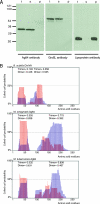
Oligomerization of mycobacterial antigen 84. (A) Subcellular localization of Ag84 in M. bovis BCG lysates. Bacterial cultures were grown at 37°C in 7H9-OADC medium, and cellular fractionation was carried out as described in Materials and Methods. Ag84 was detected in subcellular fractions by Western blot analysis using F126-2 antibody. GroEL- and lipoprotein-specific antibodies were used as controls for cytosolic and membrane proteins, respectively (t, total cell extract; s, supernatant; p, pellet). (B) Prediction of coiled-coil domains in DivIVA and Ag84 proteins. Protein sequences were analyzed with the MultiCoil (67) parallel coiled-coil prediction algorithm, using a 0.5 cutoff for the maximum scoring residue. The graphs show the calculated probabilities (y axis) for trimeric (blue bars) and dimeric (red bars) coiled coils versus the amino acid residue position (x axis). The overall probability of a coiled-coil domain is divided into a dimeric and a trimeric portion (values indicated above each domain). (C) Cross-linking of M. bovis BCG Ag84 in cell extracts (upper left panel) and purified M. bovis BCG Ag84 protein (upper right panel) by incubation with different concentrations (0.1 to 2%) of formaldehyde followed by SDS-PAGE and immunoblotting to detect mono- and oligomers with Ag84-specific F126-2 antibody. A lipoprotein-specific antibody was used as a control (lower panel). (D) Size-exclusion chromatography to analyze the molecular weight (MW) of Ag84 oligomers. Cell lysate of M. bovis BCG was applied to a gel filtration column equilibrated in PBS, and fractions were collected. Values representing elution of protein standards are indicated above the chromatogram. (E) Aliquots of the fractions indicated were loaded on 12.5% SDS gels and analyzed for the presence of Ag84 by Western blotting. Ag84 was detected in a single fraction that corresponded to a molecular weight of ∼670. The sizes of molecular weight markers used for Western blots (A, C, and E) or gel filtration (D) are indicated.
FIG. 6.
FIG. 6.
Oligomerization of mycobacterial antigen 84. (A) Subcellular localization of Ag84 in M. bovis BCG lysates. Bacterial cultures were grown at 37°C in 7H9-OADC medium, and cellular fractionation was carried out as described in Materials and Methods. Ag84 was detected in subcellular fractions by Western blot analysis using F126-2 antibody. GroEL- and lipoprotein-specific antibodies were used as controls for cytosolic and membrane proteins, respectively (t, total cell extract; s, supernatant; p, pellet). (B) Prediction of coiled-coil domains in DivIVA and Ag84 proteins. Protein sequences were analyzed with the MultiCoil (67) parallel coiled-coil prediction algorithm, using a 0.5 cutoff for the maximum scoring residue. The graphs show the calculated probabilities (y axis) for trimeric (blue bars) and dimeric (red bars) coiled coils versus the amino acid residue position (x axis). The overall probability of a coiled-coil domain is divided into a dimeric and a trimeric portion (values indicated above each domain). (C) Cross-linking of M. bovis BCG Ag84 in cell extracts (upper left panel) and purified M. bovis BCG Ag84 protein (upper right panel) by incubation with different concentrations (0.1 to 2%) of formaldehyde followed by SDS-PAGE and immunoblotting to detect mono- and oligomers with Ag84-specific F126-2 antibody. A lipoprotein-specific antibody was used as a control (lower panel). (D) Size-exclusion chromatography to analyze the molecular weight (MW) of Ag84 oligomers. Cell lysate of M. bovis BCG was applied to a gel filtration column equilibrated in PBS, and fractions were collected. Values representing elution of protein standards are indicated above the chromatogram. (E) Aliquots of the fractions indicated were loaded on 12.5% SDS gels and analyzed for the presence of Ag84 by Western blotting. Ag84 was detected in a single fraction that corresponded to a molecular weight of ∼670. The sizes of molecular weight markers used for Western blots (A, C, and E) or gel filtration (D) are indicated.
FIG. 7.
FIG. 7.
A model rationalizing how overexpression of Ag84 in M. smegmatis results in pleiomorphism in a subpopulation of cells. Overexpressed Ag84 (green) progressively accumulates at one pole (A and B). This results in cell wall expansion (red) and a localized increase in cell diameter at one pole, with septation observed at the opposite pole (C). This creates bowling-pin-shaped giant cells (D2) and small cells (D3; see Fig. 3C). Alternatively, a recovery in balanced distribution of DivIVA creates rod-shaped giant cells (D1). Untargeted accumulation of Ag84 along the lateral cell wall stimulates cell wall expansion leading to the formation of branches (D4).

Similar articles

Cited by

References

    1. Balasubramanian, M. K., D. M. Helfman, and S. M. Hemmingsen. 1992. A new tropomyosin essential for cytokinesis in the fission yeast S. pombe. Nature 360:84-87. - PubMed
    1. Balasubramanian, V., M. S. Pavelka, Jr., S. S. Bardarov, J. Martin, T. R. Weisbrod, R. A. McAdam, B. R. Bloom, and W. R. Jacobs, Jr. 1996. Allelic exchange in Mycobacterium tuberculosis with long linear recombination substrates. J. Bacteriol. 178:273-279. - PMC - PubMed
    1. Bardarov, S., S. Bardarov, Jr., M. S. Pavelka, Jr., V. Sambandamurthy, M. Larsen, J. Tufariello, J. Chan, G. Hatfull, and W. R. Jacobs, Jr. 2002. Specialized transduction: an efficient method for generating marked and unmarked targeted gene disruptions in Mycobacterium tuberculosis, M. bovis BCG and M. smegmatis. Microbiology 148:3007-3017. - PubMed
    1. Bernhardt, T. G., and P. A. de Boer. 2005. SlmA, a nucleoid-associated, FtsZ binding protein required for blocking septal ring assembly over chromosomes in E. coli. Mol. Cell 18:555-564. - PMC - PubMed
    1. Burnette, W. N. 1981. “Western blotting”: electrophoretic transfer of proteins from sodium dodecyl sulfate-polyacrylamide gels to unmodified nitrocellulose and radiographic detection with antibody and radioiodinated protein A. Anal. Biochem. 112:195-203. - PubMed

Publication types

LinkOut - more resources