. 2007 Sep 11;104(37):14640-5.
doi: 10.1073/pnas.0702426104. Epub 2007 Aug 31.

Crystal structure of human mitoNEET reveals distinct groups of iron sulfur proteins

Affiliations

Crystal structure of human mitoNEET reveals distinct groups of iron sulfur proteins

Jinzhong Lin et al. Proc Natl Acad Sci U S A. .

Abstract

MitoNEET is a protein of unknown function present in the mitochondrial membrane that was recently shown to bind specifically the antidiabetic drug pioglizatone. Here, we report the crystal structure of the soluble domain (residues 32-108) of human mitoNEET at 1.8-A resolution. The structure reveals an intertwined homodimer, and each subunit was observed to bind a [2Fe-2S] cluster. The [2Fe-2S] ligation pattern of three cysteines and one histidine differs from the known pattern of four cysteines in most cases or two cysteines and two histidines as observed in Rieske proteins. The [2Fe-2S] cluster is packed in a modular structure formed by 17 consecutive residues. The cluster-binding motif is conserved in at least seven distinct groups of proteins from bacteria, archaea, and eukaryotes, which show a consensus sequence of (hb)-C-X(1)-C-X(2)-(S/T)-X(3)-P-(hb)-C-D-X(2)-H, where hb represents a hydrophobic residue; we term this a CCCH-type [2Fe-2S] binding motif. The nine conserved residues in the motif contribute to iron ligation and structure stabilization. UV-visible absorption spectra indicated that mitoNEET can exist in oxidized and reduced states. Our study suggests an electron transfer function for mitoNEET and for other proteins containing the CCCH motif.


Conflict of interest statement

The authors declare no conflict of interest.

Figures

Fig. 1.
Fig. 1.
Multiple sequence alignment of mitoNEET homologs. Aligned are sequences from Homo sapiens (1, NP_060934; 2, NP_001008389), Mus musculus (1, NP_598768; 2, not shown), Danio rerio (1, NP_956899; 2, NP_956677), Drosophila melanogaster (NP_651684), Caenorhabditis elegans (NP_0010022387), and Arabidopsis thaliana (NP_568764). The first sequence is mitoNEET used in this study. Secondary structure elements observed in the crystal structure and a predicted transmembrane helix (TM) are shown at the top. Shown are residues whose surface area is buried by at least 30 Å2 (●) and 10 Å2 (○) due to dimerization. Asterisks denote iron-coordinating ligands. Residues with 100% and 70% conservation are shaded by dark and light green colors, respectively.
Fig. 2.
Fig. 2.
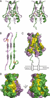
Crystal structure of the soluble domain of human mitoNEET. (A) Crosseye stereoview of the ribbon representation of the structure. The two monomer subunits are colored green and violet, respectively. The [2Fe-2S] cluster and ligand residues are represented as balls and sticks. Iron and sulfur atoms are colored orange and yellow, respectively. The secondary structure elements and N and C termini are labeled, with prime denoting the violet subunit. (B) Topology diagram of the intertwined dimer. Cysteine and histidine ligands are shown as orange and blue spots, and [2Fe-2S] clusters are shown as red rhombi. (C) The dimerization interface with the violet subunit is shown as surface representation, and the green subunit is shown as ribbons. The most highly conserved residues are colored in yellow in the violet subunit. Also shown is the relative orientation of the membrane. (D) The most highly conserved residues on the molecular surface are shown as yellow patches.
Fig. 3.
Fig. 3.
Structure of the [2Fe-2S] binding module in crosseye stereoview. (A) Stick and ball representation of the module. Backbone bonds are colored green, side chains of the core residues are pink, and side chains of noncore residues are gray. Nitrogen atoms are colored blue, oxygen atoms are colored red, sulfur atoms are colored yellow, and iron atoms are colored orange. Dashes stand for hydrogen bonds. (B) Interaction of the noncore residues with the structural environment. The two monomer subunits are colored green and violet, respectively.
Fig. 4.
Fig. 4.
The seven groups of CCCH motif-containing proteins. The 17-residue [2Fe-2S] binding motif sequences are aligned with the universally conserved core residues shaded in red, and the noncore residues that are conserved only within an individual group are shaded in blue. The domain arrangements of each group are indicated with the CCCH motif as a magenta box. Group 7 has a variable domain arrangement. Each sequence is indicated by the name of species from which it originates. The numbers of the starting and ending residues are indicated, as are the numbers of omitted residues between tandem CCCH repeats. The accession numbers of the sequences in the order of the alignment are as follows: NP_060934, NP_001022387, NP_651684, NP_956677, and NP_568764 in group 1; NP_473158, XP_669761, XP_730809, AAF99457, and XP_764182 in group 2; NP_393562, ZP_01600028, YP_658160, and NP_110689 in group 3; NP_828371, YP_956561, YP_586540, YP_005939, and ZP_00619216 in group 4; ZP_01083369, ZP_01074254, ZP_00837134, ZP_01015104, and ZP_01446837 in group 5; EAW60525, NP_497419, ZP_01694071, ZP_01167744, and NP_6101234 in group 6; and YP_565248, YP_516858, NP_275341, NP_105478, and ZP_01452291 in group 7.

References

    1. Leahy JL. Arch Med Res. 2005;36:197–209. - PubMed
    1. Yki-Jarvinen H. N EnglJ Med. 2004;351:1106–1118. - PubMed
    1. Semple RK, Chatterjee VK, O'Rahilly S. J Clin Invest. 2006;116:581–589. - PMC - PubMed
    1. Furnsinn C, Waldhausl W. Diabetologia. 2002;45:1211–1223. - PubMed
    1. Feinstein DL, Spagnolo A, Akar C, Weinberg G, Murphy P, Gavrilyuk V, Dello Russo C. Biochem Pharmacol. 2005;70:177–188. - PubMed

Publication types

MeSH terms

Associated data