. 2007 Oct;39(10):1225-34.
doi: 10.1038/ng2112. Epub 2007 Sep 2.

Modulation of morphogenesis by noncanonical Wnt signaling requires ATF/CREB family-mediated transcriptional activation of TGFbeta2

Affiliations

Modulation of morphogenesis by noncanonical Wnt signaling requires ATF/CREB family-mediated transcriptional activation of TGFbeta2

Wenlai Zhou et al. Nat Genet. 2007 Oct.

Abstract

Transcriptional readout downstream of canonical Wnt signaling is known to be mediated by beta-catenin activation of well-described targets, but potential transcriptional readout in response to noncanonical Wnt signaling remains poorly understood. Here, we define a transcriptional pathway important in noncanonical Wnt signaling. We have found that Wnt11 is a direct target of a canonical beta-catenin pathway in developing heart and that Wnt11 mutants show cardiac outflow tract defects. We provide genetic and biochemical evidence thatWnt11 signaling affects extracellular matrix composition, cytoskeletal rearrangements and polarized cell movement required for morphogenesis of the cardiac outflow tract. Notably, transforming growth factor beta2 (TGFbeta2), a key effector of organ morphogenesis, is regulated by Wnt11-mediated noncanonical signaling in developing heart and somites via one or more activating transcription factor (ATF)/cyclic AMP response element binding protein (CREB) family members. Thus, we propose that transcriptional readout mediated at least in part by a Wnt11 --> ATF/CREB --> TGFbeta2 pathway is critical in regulating morphogenesis in response to noncanonical Wnt signaling.


Figures

Figure 1
Figure 1
Wnt11 is a downstream target of Pitx2 and β-catenin in muscle cell lineages and developing heart. (a) Real-time RT-PCR analysis of Pitx2 and Wnt11 transcripts in C2C12 cells transfected with pSUPER-Pitx2 or control vector (mean ± s.d. from three independent experiments). Pitx2 and Wnt11 levels were normalized to Hprt1. (b) Top: schematic of conserved Pitx2 binding site ~850 bp upstream of the transcription start site in the Wnt11 promoter. RE, response element. Bottom: ChIP analysis using an anti-Pitx2 showed that Pitx2 was specifically recruited to the region containing the Pitx2 consensus site (primers A/B) but not to the 10-kb upstream region (primers C/D). (c) Reporter assay using a 2-kb Wnt11 promoter-reporter with or without a Pitx2 binding site mutation in C2C12 cells. Cells were transfected with either Pitx2a or Pitx2c expression vector. Error bars represent s.d. (d) Whole-mount and section RNA in situ analysis of Wnt11 expression in Pitx2-null embryos and wild-type littermates. (e) RT-PCR analysis of Wnt11 expression in C2C12 cells after lithium treatment. (f) The Wnt11 promoter was activated by canonical Wnt signaling dependent on Pitx2 binding. C2C12 cells were transfected with reporter and siRNAs as indicated and then treated with Wnt3A or LiCl before the luciferase reporter assay. Error bars represent s.d. (g) ChIP analysis of C2C12 cells in the absence or presence of both LiCl and serum using antibodies as indicated. (h) ChIP analysis of extracts from E9.5 embryonic heart. PCR was performed using primer pairs A/B and C/D.
Figure 2
Figure 2
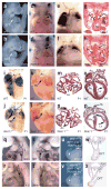
Abnormal outflow tract and great vessel development in Wnt11−/− mutants. (a,b) Right lateral view of a wild-type embryo with normal outflow tract morphology (a, arrow) and a mutant with aberrant outflow tract (b, arrow) at E9.5. (c,d) Frontal view of a wild-type embryo with normal outflow tract morphology (c, arrow) and a mutant with aberrant outflow tract (d, arrow) at E11.5. (e,f) Frontal view of wild-type (e) and mutant (f) embryos at E12.5. Wild-type embryos show an aorta displaced dorsally and pulmonary artery displaced ventrally (to the front); in mutant embryos, relative placement of the great vessels is altered. (g,h) Transverse sections of embryos in e and f. (i,j) Frontal whole-mount view of postnatal day 1 (P1) wild-type (i) and mutant (j) hearts injected with plastic dyes. In wild-type hearts, liquid blue plastic injected into the right ventricle enters the PA (i), and yellow plastic injected into the left ventricle enters the aorta (i). In mutant hearts, blue plastic injected into the right ventricle enters the aorta, and subsequently the PA; yellow plastic injected into the left ventricle enters the PA (j), showing that the aorta is connected to the right ventricle and the PA is connected to the left ventricle in the Wnt11 mutant (i.e., transposition of the great arteries (TGA, j), accompanied by a ventricular septal defect (VSD). (k,l) Whole-mount view of P1 wild-type (k) and another Wnt11 mutant heart (l), with different configuration of great vessels. (m,n) Transverse sections of wild-type (m) and mutant heart (n) show altered configuration of the great vessels: the aorta is connected to the right ventricle, and the PA is connected to the left ventricle in mutant hearts (l,n) (i.e., TGA). (o,p) Frontal sections of wild-type (o) and a different Wnt11 mutant heart at P1. This mutant shows a single outflow tract vessel which is persistent truncus arteriosus (PTA) and also had a VSD (p, arrow), with a thin-walled ventricular myocardium (p, long small arrow). (qt) Ink injection shows PAA defects in Wnt11 mutants, with smaller left 3rd PAA (r, arrow) and no apparent right 3rd PAA (t, arrow) at E9.5. (u,v) Lineage tracing of neural crest cells by Wnt1-Cre; Rosa 26 R-floxed-lacZ in Wnt11 mutant and wild-type background. (w,x) Transverse sections of embryos in u and v. OFT, outflow tract; PA, pulmonary artery; Ao, aorta; Rv, right ventricle; Lv, left ventricle; TA, truncus arteriosus; PAA, pharyngeal arch artery.
Figure 3
Figure 3
Tgfb2 as a target of the Pitx2→Wnt11 signaling pathway. (ad) Whole-mount RNA in situ hybridization with probes for Tgfb2 in wild-type (WT) (a,c) and Wnt11 mutants (b,d) at E9.5–E10.5. Tgfb2 is downregulated in Wnt11 mutants by E9.5 and at E10.5 in pharyngeal splanchnic mesoderm (white arrowhead), outflow tract (arrow) and somites (black arrowhead). (e,f) Section analysis of E9.5 embryos shows downregulation of Tgfb2 mRNA in pharyngeal mesoderm, outflow tract myocardium and endocardium in Wnt11 mutants (f) relative to wild-type littermates (e). (g,h) Section analysis of E9.5 embryos shows downregulation of Tgfb2 mRNA in somitic mesoderm (arrow) in Wnt11 mutants (h) relative to wild-type littermates (g). (i,j) Whole-mount RNA in situ hybridization with probes for Tgfb2 in wild-type (i) and Pitx2 mutants (j) at E9.75. Tgfb2 expression is decreased in pharyngeal splanchnic mesoderm (arrowhead) and outflow tract (arrow) of Pitx2 mutant embryos relative to controls. Section analysis (k,l) shows downregulation of Tgfb2 in pharyngeal mesoderm, outflow tract myocardium and endocardium of Pitx2 mutants. (m,n) Whole-mount RNA in situ hybridization with probes for Tgfb2 in wild-type embryos (m) and Vangl2 mutants (n). Vangl2 mutants show down-regulation of Tgfb2 in pharyngeal splanchnic mesoderm (black arrowhead), outflow tract (arrow) and somites (white arrowhead) (o,p). Whole-mount RNA in situ with probes for Hspg2 at E10.5. Outflow tract expression of Hspg2 is severely downregulated in Wnt11 mutants (p) relative to control littermates (o).
Figure 4
Figure 4
Tgfb2 is regulated by a Wnt11→JNK→ATF/CREB pathway. (a) Real-time RT-PCR analysis of Tgfb2 mRNA in C2C12 cells treated with Wnt11-conditioned medium (CM). Tgfb2 mRNA expression is induced by 60 min of treatment with Wnt11 CM; this induction is inhibited by treatment with SP600125, a JNK inhibitor. Tgfb2 levels were normalized to Hprt1; data represent mean ± s.d. from three independent experiments. (b) Protein blot of extracts from C2C12 cells treated with control medium (M) or Wnt11 CM. β-tubulin was a loading control. (c) Wnt11 activation of the Tgfb2 promoter depends on ATF2. C2C12 cells were transfected with a Tgfb2 promoter- reporter with or without an ATF2 binding site mutation and siRNA directed against either ATF2 or β-catenin. Cells were then treated with control medium (M) or Wnt11 CM before the luciferase assay. Error bars represent s.d. (d) ChIP analysis of C2C12 cells treated with control medium or Wnt11 CM using antibodies as indicated. (e) ChIP analysis of E9.5 heart extracts using antibodies as indicated. (f) ChIP analysis of E9.5 heart extracts from either control wild-type littermates or Wnt11 mutant embryos. (g) Expression of the Tgfb2 prom.LacZ or Tgfb2 prom.(mATF2).LacZ constructs in transgenic mouse embryos. LacZ is expressed in pharyngeal splanchnic mesoderm (arrowheads), outflow tract (white arrows) and somites (black arrows). (h) Whole-mount RNA in situ hybridization with probes for Tgfb2 in wild-type and dominant-negative ATF2 transgenic embryos. Tgfb2 expression is decreased in pharyngeal splanchnic mesoderm (arrowheads), outflow tract (white arrows) and somites (black arrows) in dominant-negative ATF2 transgenic embryos. Ace, acetylated.
Figure 5
Figure 5
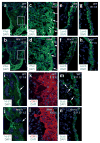
Immunofluorescence analysis of sections from wild-type and Wnt11 and Tgfb2 mutant embryos. Sections were labeled with antibodies as indicated. Anti-laminin α-5 (Lam5), anti-Scribble (Scrib), anti-integrin-β1 (Int-β1) and anti-α-smooth muscle actin (αSMA) were used. (ad) Phalloidin staining demonstrated a decrease in cytoskeletal actin in myocardial wall of the outflow tract in Wnt11 mutants. c and d show higher-magnification views of outlined areas in a and b. In e–n, views shown are the left wall of the upper outflow tract (part of the inner curvature). (e–h) Laminin α-5 shows strong and asymmetric expression at the interface between the myocardium and mesenchyme of the wild-type outflow tract (e,g). In both Wnt11 and Tgfb2 mutants, the strength of laminin α-5 expression and its ordered asymmetrical distribution are reduced (f,h). (i,j) Immunostaining for integrin β1, a marker of basal lamina, demonstrated a clear basal lamina structure in the myocardial epithelium of the wild-type outflow tract (i). In contrast, integrin β1 appeared to be severely diminished or absent in Wnt11 mutants (j), suggesting a disruption of basal structures. (k,l) Staining with antibody to αSMA demonstrated the loose mesenchymal appearance of myocardial cells extending filopodia toward the cushion mesenchyme of the wild-type outflow tract at E12.5 (k). In contrast, in Wnt11 mutants demonstrated a more compact, tightly adhering group of myocardial cells, with few filopodia (l). (m,n) Scribble protein showed a punctuate perimembranous distribution in wild-type outflow tract myocardium at E11.5, with scattered cells within the cushion mesenchyme expressing very high levels of Scribble (m). In Wnt11 mutants, there was a severe reduction in Scribble expression within the outflow tract myocardium, and no expression was observed within cushion mesenchyme cells (n).
Figure 6
Figure 6
Immunofluorescence analysis of endothelial cells from wild-type and Wnt11 mutant embryos. (af) Immunostaining of endocardial cells using VE-cadherin and PECAM-1 antibodies. Note close apposition of two endothelial layers lining the outflow tract in wild-type embryos at E12 (a,e). In Wnt11 mutants, in contrast, the two endocardial layers were not closely apposed (b,f). (c,d) Higher-magnification views of outlined areas in a and b. (g,h) Frontal sections of E14.5 wild-type and Wnt11 mutant hearts immunostaining with anti-αSMA. αSMA is expressed in the proximal part of the outflow tract cushions, where they converge with the ventricular septum (g, arrowhead), as part of the process of myocardialization. No smooth muscle actin staining is observed in this region in Wnt11 mutants (h, arrowhead). Smooth muscle actin is expressed by cardiac neural crest cells that surround the great vessels in wild-type embryos (g, arrow), and this expression is also observed in Wnt11 mutants (h, arrow), suggesting that cardiac neural crest migration and differentiation have occurred appropriately.

Similar articles

Cited by

References

    1. Veeman MT, Axelrod JD, Moon RT. A second canon. Functions and mechanisms of β-catenin-independent Wnt signaling. Dev Cell. 2003;5:367–377. - PubMed
    1. Henderson DJ, et al. Cardiovascular defects associated with abnormalities in midline development in the Loop-tail mouse mutant. Circ Res. 2001;89:6–12. - PubMed
    1. Phillips HM, Murdoch JN, Chaudhry B, Copp AJ, Henderson DJ. Vangl2 acts via RhoA signaling to regulate polarized cell movements during development of the proximal outflow tract. Circ Res. 2005;96:292–299. - PubMed
    1. Moon RT. Wnt/β-catenin pathway. Sci STKE. 2005;2005:cm1. - PubMed
    1. Chien KR. Myocyte survival pathways and cardiomyopathy: implications for trastuzumab cardiotoxicity. Semin Oncol. 2000;27:9–14. - PubMed

Publication types

MeSH terms

Associated data