Comparative Study
. 2007 Sep 3:8:39.
doi: 10.1186/1471-2121-8-39.

Fluorescence staining of live cyanobacterial cells suggest non-stringent chromosome segregation and absence of a connection between cytoplasmic and thylakoid membranes

Affiliations
Comparative Study

Fluorescence staining of live cyanobacterial cells suggest non-stringent chromosome segregation and absence of a connection between cytoplasmic and thylakoid membranes

Dirk Schneider et al. BMC Cell Biol. .

Abstract

Background: In spite of their abundance and importance, little is known about cyanobacterial cell biology and their cell cycle. During each cell cycle, chromosomes must be separated into future daughter cells, i.e. into both cell halves, which in many bacteria is achieved by an active machinery that operates during DNA replication. Many cyanobacteria contain multiple identical copies of the chromosome, but it is unknown how chromosomes are segregated into future daughter cells, and if an active or passive mechanism is operative. In addition to an outer and an inner cell membrane, cyanobacteria contain internal thylakoid membranes that carry the active photosynthetic machinery. It is unclear whether thylakoid membranes are invaginations of the inner cell membrane, or an independent membrane system.

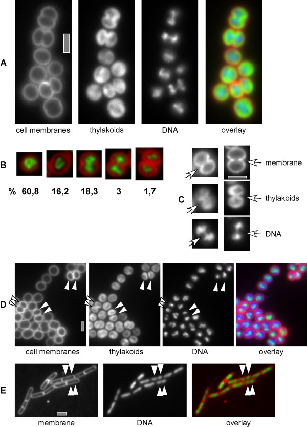
Results: We have used different fluorescent dyes to study the organization of chromosomes and of cell and thylakoid membranes in live cyanobacterial cells. FM1-43 stained the outer and inner cytoplasmic membranes but did not enter the interior of the cell. In contrast, thylakoid membranes in unicellular Synechocystis cells became visible through a membrane-permeable stain only. Furthermore, continuous supply of the fluorescent dye FM1-43 resulted in the formation of one to four intracellular fluorescent structures in Synechocystis cells, within occurred within 30 to 60 minutes, and may represent membrane vesicles. Using fluorescent DNA stains, we found that Synechocystis genomic DNA is compacted in the cell centre that is devoid of thylakoid membranes. Nucleoids segregated very late in the cell cycle, just before complete closing of the division septum. In striking contrast to Bacillus subtilis, which possesses an active chromosome segregation machinery, fluorescence intensity of stained nucleoids differed considerably between the two Synechocystis daughter cells soon after cell division.

Conclusion: Our experiments strongly support the idea that the cytoplasmic and thylakoid membranes are not directly connected, but separate entities, in unicellular cyanobacteria. Our findings suggest that a transport system may exist between the cytoplasmic membrane and thylakoids, which could mediate the extension of thylakoid membranes and possibly also protein transport from the cytoplasmic membrane to thylakoid membranes. The cell cycle studies in Synechocystis sp. PCC 6803 show that the multiple chromosome copies per cell segregate very late in the cell cycle and in a much less stringent manner than in B. subtilis cells, indicating that chromosomes may become segregated randomly and in a passive fashion, possibly through constriction of the division septum.


Figures

Figure 1
Figure 1
Fluorescence microscopy of exponentially grown Synechocystis cells. A) cells in the blue channel, right panel is scaled to the middle (blue) panel in E, B) cells in the green channel, right panel is scaled to the right (green) channel in C and to D, C) cells in the red channel (left panel, autofluorescence) and in the green channel after addition of FM1-43 membrane stain. D) cells in the green channel (upper panel) after addition of mitotracker membrane stain, or in the red channel (autofluorescence of proteins within thylacoids). E) FM1-43 membrane stain (upper panel), DAPI DNA stain (middle panel) and thylakoid autofluorescence (lower panel), grey triangles indicate division septa between separated thylakoids, F) FM1-43 stain and thylakoid autofluorescence, white triangles indicate outer membranes that have completely separated daughter cells, G) FM1-43 stain of cells subjected to conditions inducing plasmolysis (note that this is a composite image), H) Fischerella sp. cells stained with FM1-43 (green channel), white triangles indicate septa between cells. Grey bars 2 μm.
Figure 2
Figure 2
Continuous addition of FM1-43 to growing Synechocystis cells. A) Time course of cells incubated on FM1-43 containing agarose, white triangles indicate arising patches of FM1-43, grey triangle a moving FM1-43 patch. B) Z-series through cells after incubation on FM1-43 containing agarose for 45 min, position within the focal plane is indicated above/below the images. Grey bars 2 μm.
Figure 3
Figure 3
Cell cycle of Synechocystis. A) Growing cells stained with DAPI (DNA) and FM1-43 (membranes), overlay of membranes (red), thylakoids (green) and DNA (blue). B) Overlay of membrane (red) and DNA (green) of representative cells of the 5 classes defined in the text. Numbers indicate the percentage of cells present in a certain stage in an exponentially growing culture C) Fine structure of DNA (DAPI) structure in cells with a deep membrane (FM1-43) constriction and non-separated thylakoids (autofluorescence). Arrows point out non-separated nucleoids at the division plane. D) Exponentially growing cells stained with FM1-43 (membrane), DAPI (DNA), white triangles point out completely separated nucleoids having considerably different staining intensity, boxed triangles point out roughly equal nucleoids. E) Exponentially growing Bacillus subtilis cells, membrane and DNA stain analogous to panels A-D, white triangles indicate separated nucleoids in cells that do not yet have a visible division septum. Grey bars 2 μm.
Figure 4
Figure 4
Distribution of the relative DNA content of daughter cells of dividing Synechocystis cells based on DAPI stains. Of two daughter cells the smaller DNA content was each divided by the larger one, the ratios are represented by the grey bars. In Bacillus the relative nucleoid ratios varied between 0.76–1 with a mean of 0.94 and a standard deviation of 0.05. In Synechocystis the relative ratio scattered from 0.4–1 with a mean of 0.85 and a standard deviation of 0.13.

Similar articles

Cited by

References

    1. Giraldo R. Common domains in the initiators of DNA replication in Bacteria, Archaea and Eukarya: combined structural, functional and phylogenetic perspectives. FEMS Microbiol Rev. 2003;26:533–554. doi: 10.1111/j.1574-6976.2003.tb00629.x. - DOI - PubMed
    1. Lutkenhaus J. The regulation of bacterial cell division: a time and place for it. Curr Opinion Microbiol. 1998;1:210–215. doi: 10.1016/S1369-5274(98)80013-1. - DOI - PubMed
    1. Wu LJ. Structure and segregation of the bacterial nucleoid. Curr Opin Genet Dev. 2004;14:126–132. doi: 10.1016/j.gde.2004.01.006. - DOI - PubMed
    1. Volkov AV, Mascarenhas J, Andrei-Selmer C, Ulrich HD, Grauman PL. A prokaryotic condensin/cohesin-like complex can actively compact chromosomes from a single position on the nucleoid. Mol Cell Biol. 2003;23:5638–5650. doi: 10.1128/MCB.23.16.5638-5650.2003. - DOI - PMC - PubMed
    1. Defeu Soufo HJ, Graumann PL. Dynamic movement of actin-like proteins within bacterial cells. EMBO Reports. 2004;5:789–794. doi: 10.1038/sj.embor.7400209. - DOI - PMC - PubMed

Publication types

LinkOut - more resources