

Transnasal Transsphenoidal Endoscopic Repair of CSF Leakage Using Multilayer Acellular Dermis
- PMID: 17768442
- PMCID: PMC1876155
- DOI: 10.1055/s-2007-970556
Transnasal Transsphenoidal Endoscopic Repair of CSF Leakage Using Multilayer Acellular Dermis
Abstract
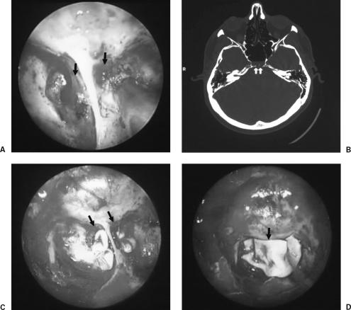
Cerebrospinal fluid (CSF) leaks result from a communication between the subarachnoid space and the upper aerodigestive tract. Because of the risk of complications such as meningitis, brain abscess, and pneumocephalus, all persistent CSF leaks should be repaired. Surgical repair may be achieved transcranially or extracranially using a wide variety of autogenous, allogenic, and synthetic patching materials. We report our results with a transnasal transsphenoidal endoscopic approach for the repair of CSF leaks coupled with a multilayer closure using acellular dermis (Allodermtrade mark). We conducted a retrospective review of all patients presenting to our institution over the past 5 years with isolated sphenoid sinus CSF fistulas.
Results: Twenty-one patients were included in the study. Nineteen patients (90.5%) had their sphenoid sinus CSF fistula repaired during the first attempt; 2 patients (9.5%) needed a second attempt. The multilayer repair of the CSF leak using acellular dermis via a transsphenoidal endoscopic approach is an effective and successful method of surgical repair of the fistula site. Neither the number, size, nor cause of the CSF fistula affected surgical outcomes. However, the presence of hydrocephalus was a significant negative variable, altering the surgical outcomes of our patients. The acellular dermis offers the advantage of not requiring autogenous tissue for the effective repair of CSF leaks in the sphenoid sinus.
Figures
References
-
- Stankiewicz J A. Cerebrospinal fluid fistula and endoscopic sinus surgery. Laryngoscope. 1991;101:250–256. - PubMed
-
- Church C A, Chiu A G, Vaughan W C. Endoscopic repair of large skull base defects after powered sinus surgery. Otolaryngol Head Neck Surg. 2003;129:204–209. - PubMed
-
- Johnson D B, Brennan P, Toland J, O'Dwyer A J. Magnetic resonance imaging in the evaluation of cerebrospinal fluid fistulae. Clin Radiol. 1996;51:837–841. - PubMed
LinkOut - more resources
Full Text Sources
Miscellaneous
