. 2007 Aug 31;3(8):e115.
doi: 10.1371/journal.ppat.0030115.

Dual targeting of antioxidant and metabolic enzymes to the mitochondrion and the apicoplast of Toxoplasma gondii

Affiliations

Dual targeting of antioxidant and metabolic enzymes to the mitochondrion and the apicoplast of Toxoplasma gondii

Paco Pino et al. PLoS Pathog. .

Abstract

Toxoplasma gondii is an aerobic protozoan parasite that possesses mitochondrial antioxidant enzymes to safely dispose of oxygen radicals generated by cellular respiration and metabolism. As with most Apicomplexans, it also harbors a chloroplast-like organelle, the apicoplast, which hosts various biosynthetic pathways and requires antioxidant protection. Most apicoplast-resident proteins are encoded in the nuclear genome and are targeted to the organelle via a bipartite N-terminal targeting sequence. We show here that two antioxidant enzymes-a superoxide dismutase (TgSOD2) and a thioredoxin-dependent peroxidase (TgTPX1/2)-and an aconitase are dually targeted to both the apicoplast and the mitochondrion of T. gondii. In the case of TgSOD2, our results indicate that a single gene product is bimodally targeted due to an inconspicuous variation within the putative signal peptide of the organellar protein, which significantly alters its subcellular localization. Dual organellar targeting of proteins might occur frequently in Apicomplexans to serve important biological functions such as antioxidant protection and carbon metabolism.


Conflict of interest statement

Competing interests. The authors have declared that no competing interests exist.

Figures

Figure 1
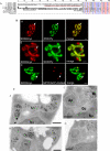
Figure 1. TgSOD2 Contains an Unusual Bipartite N-Terminal Signal and Is Targeted to both Mitochondrion and Apicoplast
(A) Alignment of the N-termini of SOD sequences from apicomplexan parasites and two bacteria. Putative SPs as predicted by SignalP are underlined in black, and putative mitochondrial transit peptides as predicted by TargetP and MitoProt are underlined in red. The arrowhead denotes the arginine residue mutated in another study [16]. The numbers in black circles above the PfSOD2 sequence denote the two first methionines. (B) Bimodal targeting of TgSOD2. Incomplete colocalization of SOD2myc with the two mitochondrial markers PRX3Ty and SOD3Ty shows that SOD2 is targeted to both the mitochondrion and an additional compartment (panels A and B). Colocalization of SOD2myc (in green) with the apicoplast marker SPTP(ACP)-DsRed demonstrates that SOD2 is also targeted to the apicoplast (panel C). Scale bars represent 2 μm. (C) Immunoelectron microscopy on parasites stably expressing tagged proteins reveals SOD2Ty to be present in both the mitochondrion and the apicoplast of T. gondii (panels A and B), whereas SOD3Ty exclusively localizes to the parasite mitochondrion (panels C and D). Panels C and D depict two serial sections of the same cell (one section in between not shown). Ap, apicoplast; Go, Golgi apparatus; Mi, mitochondrion; Nu, nucleus. The arrowheads indicate the multiple membranes surrounding the apicoplast. Scale bars represent 200 nm.
Figure 2
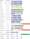
Figure 2. Overview of Recombinant Constructs Expressed in T. gondii
Various T. gondii full-length proteins and protein domains were expressed as tagged fusion proteins (employing myc- or Ty-protein tags, DsRed, and/or GFP) to determine their intracellular localization. The overview shows the name of the recombinant construct, the experimentally determined localization, and a diagrammatic representation of the fusion protein indicating the exact boundaries of protein domains (numbers refer to amino acids). If not specified further, SP, TP, and SPTP refer to the predicted signal peptide, transit peptide, and the putative bipartite leader of TgSOD2, respectively. Brackets indicate SP and TP domains taken from other proteins (SOD3 or ACP). For example, SPTP-SOD3Ty refers to the putative bipartite leader of SOD2 (amino acids 1–91; dark blue) followed by the predicted mature protein portion of SOD3 (amino acids 57–258; light blue) followed by a C-terminal Ty-tag, whereas TP(SOD3)-SOD2Ty refers to the predicted mitochondrial transit peptide of SOD3 (amino acids 1–56; light blue) followed by the predicted mature protein portion of SOD2 (amino acids 92–287; light blue) followed by a C-terminal Ty-tag.
Figure 3
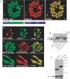
Figure 3. The N-Terminal Targeting Sequence of SOD2 Is Not Sufficient for Bimodal Targeting of Reporter Proteins
(A) The predicted bipartite leader of SOD2 (SPTP) fused to reporter proteins (GFPmyc or DsRed) only localizes to the parasite mitochondrion as revealed by colocalization with the bimodally targeted SOD2 (SOD2Ty) (panels a and b), indicating that part of the information necessary for bimodal targeting of SOD2 resides in its mature protein portion. Replacement of the GFP or DsRed reporter by the predicted mature protein of SOD3 complements the bipartite leader of SOD2 (SPTP-SOD3Ty) and restores bimodal targeting to both organelles (panel c). The putative mitochondrial transit peptide of SOD3 in conjunction with the SOD2 mature protein leads to mitochondrial localization (panel d). The arrows indicate the apicoplast. Scale bars represent 2 μm. (B) Western blot analysis of parasites stably expressing SOD2Ty, SOD3Ty, SPTP-SOD3Ty, and TP(SOD3)-SOD2Ty using anti-Ty-1 antibodies. (C) Western blot analysis of parasite clones stably expressing SOD2myc and SPTP(SOD2)-GFPmyc using anti-myc and anti-actin antibodies, showing that the bimodal targeting of SOD2myc is not due to a difference in the expression level compared with the mitochondrion-targeted SPTP-GFPmyc.
Figure 4
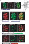
Figure 4. Dissection of the Bipartite Targeting Signal of TgSOD2
(A) Double IFA on parasites expressing stable TP-SOD2Ty and transient SOD2myc showed that removal of the SP abolishes targeting to the apicoplast, localizing the protein to the mitochondrion and the cytosol. The arrows indicate the apicoplast. Scale bars represent 2 μm. (B) Western blot analysis using anti-Ty-1 antibodies confirms incomplete targeting of TP-SOD2Ty to the organelle by showing that a fraction of the protein remains unprocessed. The putative SP of TgSOD2 acts as a suboptimal SP by only partially targeting SOD2 to the apicoplast. (C) Colocalization of parasites expressing SOD2TyHDEL and ΔTPSOD2TyHDEL. Insertion of an ER retention signal on SOD2 that lacks the TP(ΔTPSOD2TyHDEL) leads to accumulation of the protein in the ER while insertion of HDEL on SOD2 (SOD2TyHDEL) does not prevent targeting to the mitochondrion (Figure 4C). This demonstrates that SOD2 is transported independently to the two organelles. (D) When fused to the protein FNR or ACP, the SP of TgSOD2 caused a dramatic relocalization of FNR and ACP to the mitochondrion with occasional apicoplast accumulation. The arrows indicate the apicoplast.
Figure 5
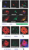
Figure 5. PfSOD2 Is Targeted to the Apicoplast in the Intra-Erythrocytic Stages of P. falciparum
(A) IFA on live erytrocytes infected with transgenic P. falciparum parasites expressing PfSOD2GFP. The construct starts either at the second methionine ( MNLKIL…) and is controlled by the PfCRT promoter (panel a), or at the first methionine (MLYKLY…) and is controlled by the PfSOD2 promoter (panel b) (see Figure 1A). The mitochondrion is labeled in red with MitoTracker and the localization PfSOD2-GFP to the apicoplast is detected in green. The same construct was stably expressed in the strain 3D7 and the same results were observed. Moreover, PfSOD2 was also shown to localize to the apicoplast only in the gametocytes (panel c). The arrows indicate the apicoplast. (B) IFA on stable transgenic T. gondii parasites expressing PfSOD2 with a Ty tag at the C-terminus. The construct starts either at the second methionine (panel a) or at the first methionine (panel b). The periphery of T. gondii parasites is labeled in red with anti-MLC1 antibodies, while the localization of the fusion protein PfSOD2-Ty to the apicoplast is shown in green.
Figure 6
Figure 6. Dual Targeting of a T. gondii TPX
(A) Schematics showing the generation of two transcript variants consisting of five exons each (E1–E5) by differential splicing and the resulting gene products TPX1/1 and TPX1/2. (B) Alignment of apicomplexan TPX sequences and related glutathione peroxidases. Putative SPs as predicted by SignalP are underlined in black (a broken line indicates a positive prediction by the Hidden Markov Models only, and not by the Neural Networks), and putative mitochondrial or chloroplast transit peptides as predicted by TargetP or MitoProt are underlined in red or green, respectively. (C) Parasites stably expressing TgTPX1/2Ty are transiently transfected with the apicoplast and mitochondrial markers, FNR-DsRed and SPTP-GFPmyc, respectively. Double IFA reveals dual targeting of TgTPX1/2Ty to both organelles. The arrows indicate the apicoplast. Scale bars represent 2 μm. (D) Western blot analysis (using anti-Ty-1 antibodies) reveals processing of TgTPX1/1Ty and TPX1/2Ty in parasites stably expressing these proteins.
Figure 7
Figure 7. Bimodal Targeting of T. gondii ACN/IRP
(A) Alignment of TgIRP/ACN with related protein sequences. Red arrowheads indicate theoretical alternative start-methionines. A putative SP as predicted by SignalP is underlined in black, and putative mitochondrial transit peptides (as predicted by TargetP or MitoProt) are underlined in red (both for the full-length sequence and for a hypothetical protein starting from the second methionine at position 74). (B) Colocalisation reveals bimodal targeting of SPTP(ACN)GFP-Ty fusion protein to the parasite mitochondrion and apicoplast. A deletion of the SP targets the fusion protein (TP(ACN)-GFPTy) to the mitochondrion only, while a mutation of the second methionine in position 74 within the TP results in bimodal targeting (SPTP(ACN)-M74I-GFP-Ty). The asterisk indicates the position of the mutation M74I. The arrows indicate the apicoplast. Scale bars represent 2 μm. (C) Colocalization of TgICDH1Ty (left panel) with the apicoplast marker FNRDsRed (middle panel) indicates a localization of ICDH1 around the periphery of the plastid organelle. Scale bars represent 2 μm.
Figure 8
Figure 8. Metabolic Maps
Overview of select metabolic pathways of T. gondii taken into consideration in this study. Illustrated is the established (in black or blue) or putative (in red) localization of enzymes and pathways involved in (i) antioxidant defense and (ii) in the mitochondrial Krebs cycle and in similar metabolic reactions in the apicoplast. Blue, organelle-specific enzymes and pathways; red, enzymes and reactions whose subcellular localization is hypothetical. IRE, iron-responsive element; PRX, peroxiredoxin; PyC, pyruvate carboxylase; TLP, thioredoxin-like protein.

Similar articles

Cited by

References

    1. Ralph SA, Van Dooren GG, Waller RF, Crawford MJ, Fraunholz MJ, et al. Tropical infectious diseases: Metabolic maps and functions of the Plasmodium falciparum apicoplast. Nat Rev Microbiol. 2004;2:203–216. - PubMed
    1. Gardner MJ, Hall N, Fung E, White O, Berriman M, et al. Genome sequence of the human malaria parasite Plasmodium falciparum . Nature. 2002;419:498–511. - PMC - PubMed
    1. Wilson RJ. Progress with parasite plastids. J Mol Biol. 2002;319:257–274. - PubMed
    1. Sato S, Clough B, Coates L, Wilson RJ. Enzymes for heme biosynthesis are found in both the mitochondrion and plastid of the malaria parasite Plasmodium falciparum . Protist. 2004;155:117–125. - PubMed
    1. Ellis KE, Clough B, Saldanha JW, Wilson RJ. Nifs and Sufs in malaria. Mol Microbiol. 2001;41:973–981. - PubMed

Publication types

LinkOut - more resources