. 2007 Aug;3(8):e139.
doi: 10.1371/journal.pgen.0030139. Epub 2007 Jul 6.

Mutation in mouse hei10, an e3 ubiquitin ligase, disrupts meiotic crossing over

Affiliations

Mutation in mouse hei10, an e3 ubiquitin ligase, disrupts meiotic crossing over

Jeremy O Ward et al. PLoS Genet. 2007 Aug.

Abstract

Crossing over during meiotic prophase I is required for sexual reproduction in mice and contributes to genome-wide genetic diversity. Here we report on the characterization of an N-ethyl-N-nitrosourea-induced, recessive allele called mei4, which causes sterility in both sexes owing to meiotic defects. In mutant spermatocytes, chromosomes fail to congress properly at the metaphase plate, leading to arrest and apoptosis before the first meiotic division. Mutant oocytes have a similar chromosomal phenotype but in vitro can undergo meiotic divisions and fertilization before arresting. During late meiotic prophase in mei4 mutant males, absence of cyclin dependent kinase 2 and mismatch repair protein association from chromosome cores is correlated with the premature separation of bivalents at diplonema owing to lack of chiasmata. We have identified the causative mutation, a transversion in the 5' splice donor site of exon 1 in the mouse ortholog of Human Enhancer of Invasion 10 (Hei10; also known as Gm288 in mouse and CCNB1IP1 in human), a putative B-type cyclin E3 ubiquitin ligase. Importantly, orthologs of Hei10 are found exclusively in deuterostomes and not in more ancestral protostomes such as yeast, worms, or flies. The cloning and characterization of the mei4 allele of Hei10 demonstrates a novel link between cell cycle regulation and mismatch repair during prophase I.


Conflict of interest statement

Competing interests. The authors have declared that no competing interests exist.

Figures

Figure 1
Figure 1. The mei4 Mutation Inhibits Normal Meiotic Chromosome Alignment at Metaphase I in Males and Females and Is Associated with Apoptosis in Spermatocytes
(A) Heterozygous (mei4/+) seminiferous tubule cross sections stained with hematoxylin and eosin show normal telophase and metaphase I structures (black arrow and arrowheads, respectively) as well as postmeiotic round spermatids and elongating differentiating spermatids (white arrows). (B) Homozygous mutant (mei4/mei4) sections show abnormal metaphase and anaphase I-like spermatocytes with disrupted chromosome congression to the metaphase plate and aberrant chromosome distribution on the spindle (black arrows). (C and D) mei4/mei4 mutant seminiferous tubules have significantly greater numbers of apoptotic cells (brown cell, black arrow) than +/+ tubules (D versus C, respectively) as determined by TUNEL assay. The average number of TUNEL positive cells per 0.146-mm2 field in tubules that contained apoptotic cells was 140 for mei4/mei4 versus 9.6 for +/+ (p = 3.28 × 10⁻¹⁵). (E and F) The ovaries of mei4/mei4 females appear histologically normal. Hematoxylin and eosin-stained cross sections reveal follicles and oocytes in all stages of development in normal (E) and mutant animals (F). (G and H) Hoechst-stained metaphase I chromosome structures occur normally in mei4/+ oocytes (white arrow, G) and abnormally (asterisks, H) in mei4/mei4 oocytes. Infrequently, normal metaphase chromosomes structures are seen in mutant oocytes (white arrow, [H]). Spindles in (G and H) are labeled with an anti-b tubulin antibody.
Figure 2
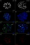
Figure 2. mei4/mei4 Spermatocytes Synapse in Prophase I but Have Few Intact Bivalents at Metaphase I
(A and B) Metaphase chromosome spreads were prepared from normal (mei4/+ [A]) and mutant (mei4/mei4 [B]) males and stained with DAPI. In (A) mei4/+ spreads exhibit the expected number of bivalent recombination structures. (B) depicts a mei4/mei4 spread with few chiasmata (white arrow) and with most chromosomes present as univalents. (C–F) Chromosome spreads from normal (+/+) and mutant (mei4/mei4) spermatocytes were labeled with antibodies to SYCP3 and counter-stained with DAPI at various stages of MI. Pachytene (C and D) +/+ and mei4/mei4 spermatocytes are similar. At diplonema, +/+ (E) spreads show pairs of homologous chromosomes associated at sites of recombination (white arrows) and or centromeres. In contrast, in mei4/mei4 spreads (F) many chromosomes are univalents associated neither at the centromere nor sites of recombination (white arrowheads). The loss of bivalent cohesion is not complete however, and some chiasmata are seen in mei4/mei4 preparations. (G and H) Chromosome spreads from heterozygote (mei4/+ [G]) and homozygous recessive (mei4/mei4 [H]) animals were labeled (red) with antibodies to SYCP1, a marker of the central element and synapsis. In both cases SYCP1 is present indicating that the transverse elements of the SC are forming and that chromosomes in mei4/mei4 spermatocytes synapse normally in pachynema.
Figure 3
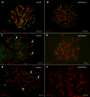
Figure 3. Chromosomes in mei4/mei4 Spermatocytes Contain RAD51 Foci in Zygonema but Lack MLH3 and MLH1 Foci in Pachynema
(A and B) The DSB repair protein RAD51 appears in foci (green) on zygotene chromosomes in both mei4/+ and mei4/mei4 chromosome spreads counter labeled with anti-SYCP3 (red). (C–F) Chromosome spreads from heterozygous (mei4/+) and mutant (mei4/mei4) spermatocytes were labeled with antibodies to MLH3 (green [C and D]) or MLH1 (green [E and F]) and counter-labeled with anti-SYCP3 (red). MLH3 and MLH1 foci (white arrows) are present on mei4/+ chromosomes (C) and (E) but not on mei4/mei4 chromosomes (D) and (F).
Figure 4
Figure 4. mei4 Maps to a 2.5-mb Region on Chromosome 14 and Is Associated with an Altered Hei10 Transcript That Is Expressed Prominently in the Testis and 17-d Embryo
(A) The diagram shows microsatellite loci abbreviated with “M” for D14Mit. To map mei4 genetically, intersubspecific intercrosses were set up between animals heterozygous for mei4. The number of meioses scored for recombinants in particular intervals are indicated in raw numbers (# of recombinants/# of meioses) and recombination fractions. The data placed mei4 in a 0.7-cM region corresponding to a physical distance of ∼2.5 mb. (B) RT-PCR was performed on total RNA isolated from normal (+/+) and mutant (mei4/mei4) animals. Neither Parp2 nor Apex1 showed altered transcript amount or size (rows 2 and 3, respectively). The Hei10 transcript is expressed at similar levels in +/+ and mei4/mei4 but displays altered mobility in agarose gel electrophoresis (row 4). Similar b-Actin (Actb) expression in both +/+ and mei4/mei4 indicates equal RNA amounts (row 1). (C) RT-PCR of +/+, mei4/+, and mei4/mei4 animals. The +/+ (lane 1) displays the expected 430-bp band, whereas the mei4/+ (lane 2) displays both the expected size band and a smaller band (358 bp) that is identical in size to the only band in the mei4/mei4 sample (lane 3). (D) A mouse cDNA panel was used to determine the expression pattern of Hei10 across a range of tissues. Expression relative to the GAPDH control was highest in testis and 17-d-old embryo. The 17-d-old embryo is a mixed male and female sample and represents the onset to midterm of prophase I in the mouse oocyte.
Figure 5
Figure 5. Genomic Sequencing of Hei10 Reveals a Mutation in the Consensus GT 5′ Splice Site in Intron 1
(A) Genomic DNA from +/+, mei4/+, and mei4/mei4 animals was sequenced, and a single base transversion (g > t, gray shading) was found in the 5′ splice site consensus GT sequence. Exon sequence is denoted with capital letters and intron sequence with lower case. (B) The 5′ splice site of intron 1 contains the consensus GT dinucleotide (Hei10). The splice site mutation described in (A) (g > t, red t indicates mutant form) causes the splicing machinery to select a GT site 72 bp upstream of the original site (dashed line). The 72-bp in-frame section of exon 1 is removed with intron 1 and is not present in the mature transcript (sequence not shown). The predicted protein sequence of HEI10 in mei4/mei4 animals contains a deletion of amino acids (a.a.) 76–100 including a conserved putative cyclin binding domain (red RAL).
Figure 6
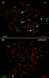
Figure 6. CDK2 Does Not Localize to Interstitial Sites on Synapsed mei4/mei4 Bivalents during Pachynema
(A) Wild-type (+/+) spermatocyte chromosome spread labeled with antibodies against CDK2 (green) and SYCP3 (red). CDK2 localizes to telomeric foci (open arrowheads), along the asynapsed axes of the sex chromosomes (closed arrowheads), and at interstitial sites (white arrows) along synapsed wild-type bivalents in pachynema. (B) mei4/mei4 spermatocyte chromosome spread labeled as in (A) showing absence of CDK2 localization at interstitial loci. Despite loss at interstitial sites, CDK2 localization is observed at telomeric foci (open arrowheads) and at loci along the sex chromosomes (closed arrowheads).
Figure 7
Figure 7. ClustalW Protein Sequence Alignment of HEI10 Orthologs from Deuterostome Lineages
Sequence from Homo sapiens (human) and Mus musculus (mouse) have been experimentally determined, while the sequences from Canis familiaris (dog), Bos taurus (cow), Monodelphis domestica (opossum), Ornithorhynchus anatinus (platypus), Xenopus tropicalis (clawed frog), Gasterosteus aculeatus (stickleback fish), Danio rerio (zebrafish), Ciona intestinalis (transparent sea squirt), and Strongylocentrotus purpuratus (California purple sea urchin) were available in databases as predicted proteins (accession numbers are listed below). A candidate RING domain, a potential RXL-type B-type cyclin interaction motif (RAL), the deleted portion of the Hei10mei4 allele (asterisks), and a central coiled-coil domain are indicated with text above the sequences. Using database and BLAST searches, HEI10 orthologs could not be identified in the chicken, yeast, or in any protostomes such as D. melanogaster or C. elegans.
Figure 8
Figure 8. Proposed Mechanism: HEI10mei4 Mediated Failure to Degrade CCNB3 Leads to Inability to Recruit MLH3 and MLH1 Resulting in Failed Recombination
(A) During normal Prophase I, subsequent to DSB repair via RAD51, HEI10 mediates the degradation of CCNB3 (B3) freeing CDK2 to associate with interstitial sites on chromosome cores in pachynema. Subsequently (or contemporaneously), MLH3 and MLH1 are recruited to recombination nodules containing CDK2. In diplonema, MMR has occurred, MLH3, MLH1, and CDK dissociate from the cores and chiasmata maintain homolog association until the onset of anaphase I. (B) In mei4/mei4 animals, DSB formation and RAD51 foci occur normally. Inability of HEI10mei4 to associate with B3 leads to the accumulation or mislocalization of B3 and the titration of the available CDK2. CDK2 is unable to associate with sites of recombination, as are MLH3 and MLH1. In our model, failure to correct mismatches during Holiday junction resolution leads to incomplete recombination intermediate resolution and arrest at the metaphase I spindle checkpoint.

Similar articles

Cited by

References

    1. Roeder GS, Bailis JM. The pachytene checkpoint. Trends Genet. 2000;16:395–403. - PubMed
    1. Morelli MA, Cohen PE. Not all germ cells are created equal: Aspects of sexual dimorphism in mammalian meiosis. Reproduction. 2005;130:761–781. - PubMed
    1. Keeney S, Baudat F, Angeles M, Zhou ZH, Copeland NG, et al. A mouse homolog of the Saccharomyces cerevisiae meiotic recombination DNA transesterase Spo11p. Genomics. 1999;61:170–182. - PubMed
    1. Romanienko PJ, Camerini-Otero RD. The mouse Spo11 gene is required for meiotic chromosome synapsis. Mol Cell. 2000;6:975–987. - PubMed
    1. Moens PB, Chen DJ, Shen Z, Kolas N, Tarsounas M, et al. Rad51 immunocytology in rat and mouse spermatocytes and oocytes. Chromosoma. 1997;106:207–215. - PubMed

Publication types

MeSH terms