

Trabecular structure quantified with the MRI-based virtual bone biopsy in postmenopausal women contributes to vertebral deformity burden independent of areal vertebral BMD
- PMID: 17784842
- PMCID: PMC2663589
- DOI: 10.1359/jbmr.070815
Trabecular structure quantified with the MRI-based virtual bone biopsy in postmenopausal women contributes to vertebral deformity burden independent of areal vertebral BMD
Abstract
In postmenopausal women with a wide range of vertebral deformities, MRI-based structural measures of topology and scale at the distal radius are shown to account for as much as 30% of vertebral deformity, independent of integral vertebral BMD.
Introduction: Trabecular bone architecture has been postulated to contribute to overall bone strength independent of vertebral BMD measured by DXA. However, there has thus far been only sparse in vivo evidence to support this hypothesis.
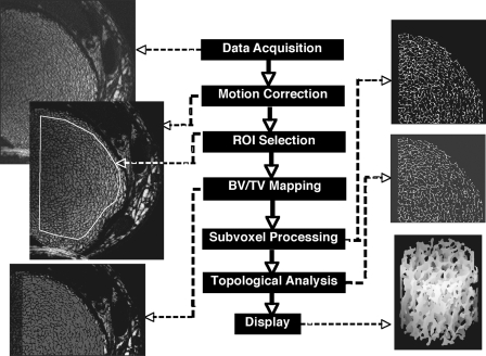
Materials and methods: Postmenopausal women, 60-80 yr of age, were screened by DXA, and those with T-scores at either the hip or spine falling within the range of -2.5 +/- 1.0 were studied with the MRI-based virtual bone biopsy, along with heel broadband ultrasound absorption and pQCT of the tibia. The data from 98 subjects meeting the enrollment criteria were subjected to microMRI at the distal tibia and radius, and measures of topology and scale of the trabecular bone network were computed. A spinal deformity index (SDI) was obtained from morphometric measurements in midline sagittal MR images of the thoracic and lumbar spine to evaluate associations between structure and deformity burden.
Results: A number of structural indices obtained at the distal radius were correlated with the SDI. Among these were the topological surface density (a measure of trabecular plates) and trabecular bone volume fraction, which were inversely correlated with SDI (p < 0.0001). Combinations of two structural parameters accounted for up to 30% of the variation in SDI (p < 0.0001) independent of spinal BMD, which was not significantly correlated. pQCT trabecular BMD was also weakly associated, whereas broadband ultrasound absorption was not. No significant association between SDI and structural indices were found at the tibia.
Conclusions: Structural measures at the distal radius obtained in vivo by microMRI explained a significant portion of the variation in total spinal deformity burden in postmenopausal women independent of areal BMD.
Figures


Similar articles
-
Trabecular architecture and vertebral fragility in osteoporosis.Curr Osteoporos Rep. 2012 Jun;10(2):132-40. doi: 10.1007/s11914-012-0097-0. Curr Osteoporos Rep. 2012. PMID: 22492119 Review.
-
Vertebral deformities and fractures are associated with MRI and pQCT measures obtained at the distal tibia and radius of postmenopausal women.Osteoporos Int. 2014 Mar;25(3):973-82. doi: 10.1007/s00198-013-2569-1. Epub 2013 Nov 13. Osteoporos Int. 2014. PMID: 24221453 Free PMC article.
-
In vivo assessment of architecture and micro-finite element analysis derived indices of mechanical properties of trabecular bone in the radius.Osteoporos Int. 2002 Jan;13(1):6-17. doi: 10.1007/s198-002-8332-0. Osteoporos Int. 2002. PMID: 11878456
-
Deterioration of trabecular plate-rod and cortical microarchitecture and reduced bone stiffness at distal radius and tibia in postmenopausal women with vertebral fractures.Bone. 2016 Jul;88:39-46. doi: 10.1016/j.bone.2016.04.003. Epub 2016 Apr 12. Bone. 2016. PMID: 27083398 Free PMC article.
-
Role of magnetic resonance for assessing structure and function of trabecular bone.Top Magn Reson Imaging. 2002 Oct;13(5):335-55. doi: 10.1097/00002142-200210000-00005. Top Magn Reson Imaging. 2002. PMID: 12464746 Review.
Cited by
-
Segmentation of the Proximal Femur from MR Images using Deep Convolutional Neural Networks.Sci Rep. 2018 Nov 7;8(1):16485. doi: 10.1038/s41598-018-34817-6. Sci Rep. 2018. PMID: 30405145 Free PMC article.
-
Bone density, geometry, microstructure, and stiffness: Relationships between peripheral and central skeletal sites assessed by DXA, HR-pQCT, and cQCT in premenopausal women.J Bone Miner Res. 2010 Oct;25(10):2229-38. doi: 10.1002/jbmr.111. J Bone Miner Res. 2010. PMID: 20499344 Free PMC article.
-
Trabecular architecture and vertebral fragility in osteoporosis.Curr Osteoporos Rep. 2012 Jun;10(2):132-40. doi: 10.1007/s11914-012-0097-0. Curr Osteoporos Rep. 2012. PMID: 22492119 Review.
-
Mechanical implications of estrogen supplementation in early postmenopausal women.J Bone Miner Res. 2010 Jun;25(6):1406-14. doi: 10.1002/jbmr.33. J Bone Miner Res. 2010. PMID: 20200948 Free PMC article.
-
Osteoprotective effect of hormone therapy on bone microarchitecture before impaired bone mineral density in ovariectomized rats.J Turk Ger Gynecol Assoc. 2012 Dec 1;13(4):261-6. doi: 10.5152/jtgga.2012.42. eCollection 2012. J Turk Ger Gynecol Assoc. 2012. PMID: 24592053 Free PMC article.
References
-
- Gordon CL, Webber CE, Nicholson PS. Relation between image-based assessment of distal radius trabecular structure and compressive strength. Can Assoc Radiol J. 1998;49:390–397. - PubMed
-
- Bousson V, Le Bras A, Roqueplan F, Kang Y, Mitton D, Kolta S, Bergot C, Skalli W, Vicaut E, Kalender W, Engelke K, Laredo JD. Volumetric quantitative computed tomography of the proximal femur: Relationships linking geometric and densitometric variables to bone strength. Role for compact bone. Osteoporos Int. 2006;17:855–864. - PubMed
-
- Siris ES, Brenneman SK, Barrett-Connor E, Miller PD, Sajjan S, Berger ML, Chen YT. The effect of age and bone mineral density on the absolute, excess, and relative risk of fracture in postmenopausal women aged 50-99: Results from the National Osteoporosis Risk Assessment (NORA) Osteoporos Int. 2006;17:565–574. - PubMed
-
- Recker RR, Barger-Lux MJ. The elusive concept of bone quality. Curr Osteopor Rep. 2004;2:97–100. - PubMed
-
- Paschalis EP, Boskey AL, Kassem M, Eriksen EF. Effect of hormone replacement therapy on bone quality in early postmenopausal women. J Bone Miner Res. 2003;18:955–959. - PubMed
Publication types
MeSH terms
Grants and funding
LinkOut - more resources
Full Text Sources
Medical
