. 2007 Oct;17(4):389-98.
doi: 10.1111/j.1750-3639.2007.00088.x. Epub 2007 Sep 4.

Presence of HSV-1 immediate early genes and clonally expanded T-cells with a memory effector phenotype in human trigeminal ganglia

Affiliations

Presence of HSV-1 immediate early genes and clonally expanded T-cells with a memory effector phenotype in human trigeminal ganglia

Tobias Derfuss et al. Brain Pathol. 2007 Oct.

Abstract

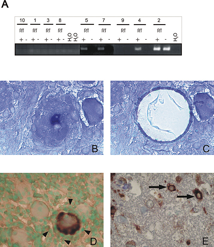
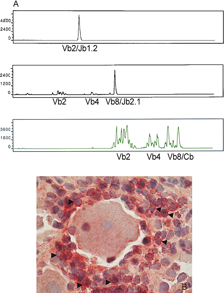
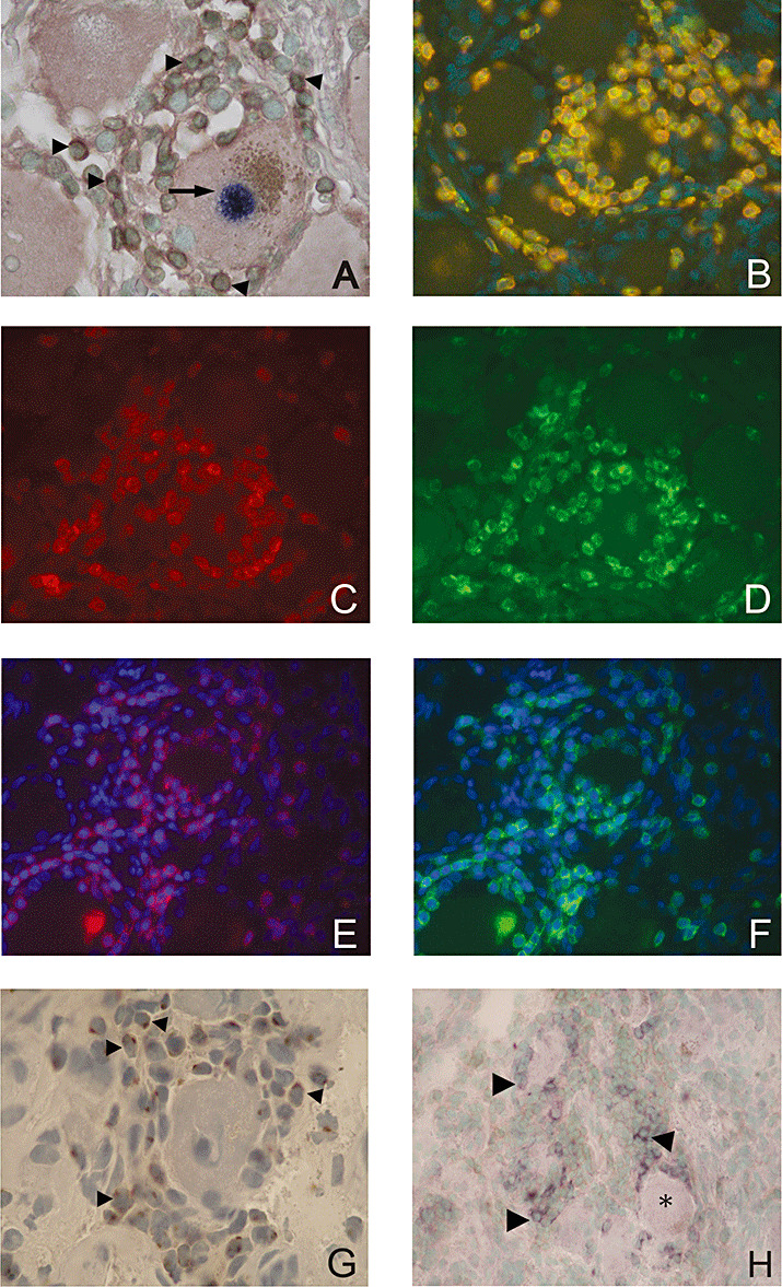
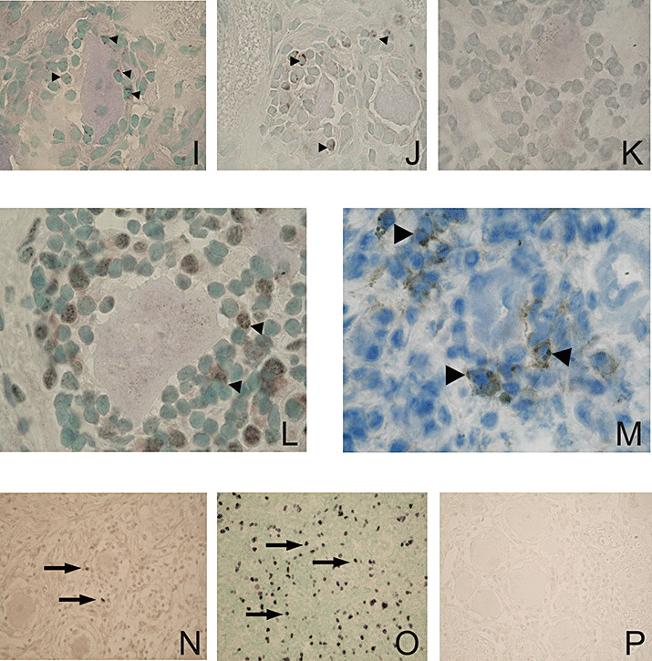
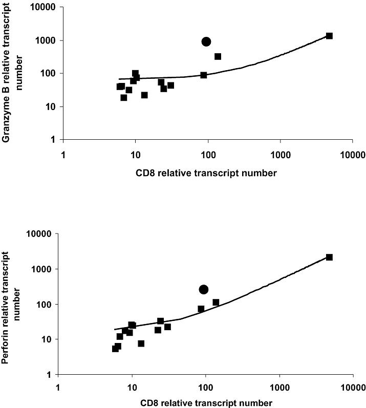
The latent persistence of herpes simplex virus type 1 (HSV-1) in human trigeminal ganglia (TG) is accompanied by a chronic CD8 T-cell infiltrate. The focus of the current work was to look for HSV-1 transcription activity as a potential trigger of the immune response and to characterize the immune cell infiltrates by this feature. We combined in situ hybridization, laser cutting microscopy, and single cell RT-PCR to demonstrate the expression of the HSV-1 immediate early (IE) genes ICP0 and ICP4 in human trigeminal neurons. Using CDR3 spectratyping, we showed that the infiltrating T-cells are clonally expanded, indicating an antigen-driven immune response. Moreover, the persisting CD8+ T-cells had features of the memory effector phenotype. The voltage-gated potassium channel Kv1.3, a marker of chronic activated memory effector cells, and the chemokines CCL5 and CXCL10 were expressed by a subpopulation of infiltrating cells. The corresponding chemokine receptors CCR5 and CXCR3 were co-expressed on virtually all CD8 T-cells. In addition, T-cells expressed granzymes and perforin. In contrast to animal models of HSV-1 latency, hardly any FoxP3-positive regulatory T-cells were detected in human TG. Thus, HSV-1 IE genes are expressed in human TG and the infiltrating T-cells bear several characteristics that suggest viral antigenic stimulation.


Figures

Figure 1
Figure 1
A. ICP4 nested RT‐PCR. The subjects shown were tested with and without reverse transcriptase (RT). A clear amplicon can be seen in the samples of subjects 4, 5 and 7 (no amplification could be seen in the corresponding samples without RT). The possibility of DNA contamination in Subject 2 cannot be excluded, as a signal is also seen in the sample without RT. BE. Laser‐cutting microdissection (LCM) and detection of ICP0 and LAT by in situ hybridization in human TG latently infected with HSV‐1; (B) LAT ISH: positive neuron shows dark blue nucleus (before LCM). C demonstrates the same section as in B after LCM; the LAT‐positive neuron has been excised. (D) ICP0 ISH: positive neuron shows dark brown cytoplasm and is surrounded by mononuclear cells (arrowheads) in the TG, counterstained with methylgreen. (E) Positive ICP0 signal in neurons of an HSV‐1 encephalitis control case (arrows). ISH = in situ hybridization; LAT = latency‐associated transcripts; RT‐PCR = reverse transcription polymerase chain reaction.
Figure 2
Figure 2
Examples of CDR3 spectratyping (A) and Vb2 IHC (B) on the same TG of Subject 1. A. In the first panel, the expansion of T‐cells with Vb2 and Jb1.2 is shown. In the second panel the expansion of T‐cells with Vb8 and Jb2.1 is provided. The sequences of these peaks are shown in Table 4A. In the third panel a polyclonal Gaussian‐like distribution of TCR lengths for Vb2, 4, 8 and Cb is shown. B. IHC with an antibody specific for Vb2 in the TG section of Subject 1 is shown. Vb2‐positive T‐cells (red cytoplasm, AEC, some labeled with arrowheads) around a neuron (×1000; counterstained with hematoxylin). CDR3 spectratyping showed an expansion of T‐cells with Vb2 in the same individual (see first panel). IHC = immunohistochemistry; TCR = T‐cell receptor; TG = trigeminal ganglia.
Figure 3
Figure 3
Characterization of the T‐cell phenotype in human TG latently infected with HSV‐1. A. Combination of LAT ISH and CD3 IHC: a LAT‐positive neuron with a dark blue nucleus (arrow) encircled by brown (diaminobenzidine) stained CD3+ T‐cells (arrowheads), counterstained with methyl green (×1000). B. Fluorescence double staining for CXCR3 and CD3: Double positive CXCR3 and CD3 T‐cells (yellow) using FITC‐labeled antibody (green) for CXCR3 and Cy3‐labeled antibody (red) for CD3, counterstained with DAPI (one‐channel image, ×400). C,D. Fluorescence double staining of CD8 (red) using Texas Red®‐labeled antibody (C) and CXCR3 (green) using FITC‐labeled antibody (D) (two‐channel image, ×400). E,F. Fluorescence double staining of CD8 (red) using Texas Red®‐labeled antibody (E) and CCR5 (green) using FITC‐labeled antibody (F), counterstained with DAPI (two‐channel image ×400). G. CCL5/RANTES producing cells (brown, arrowheads) around a sensory neuron, counterstained with hematoxylin (×1000). H. CXL10/IP‐10 producing cells (dark blue) among CD3+ infiltrates (arrowheads) and within the close vicinity of the sensory neurons (asterisk), counterstained with methyl green (×400). I. Granules containing granzyme B in the cytoplasm of T cells (brown, arrowheads) in close proximity to the neuron (×1000). J. Granules containing granzyme A in the cytoplasm of T cells (brown, arrowheads) around neurons (×1000). K. Negative control section applied in parallel to the granzyme A and B stainings (TG section incubated with 5% normal rabbit serum instead of the primary antibody), (×1000). L. Kv1.3 positive T‐cells (brown, arrowheads) among the infiltrates around a neuron (×1000). M. CD4 positive T‐cells among the infiltrates (brown, arrowheads), (×1000). N. Rare Foxp3 + T‐cells (brown, arrows) in the vicinity of the infiltrates (×400). O. Numerous Foxp3 + T‐cells (brown, arrows) in a human tonsil (positive control tissue), (×400). P. Negative control section applied in parallel to the FoxP3 staining (TG section incubated with 5% normal rabbit serum), (×400). DAPI = 4’,6‐Diamidino‐2‐phenylindole; HSV‐1 = herpes simplex virus type 1; IHC = immunohistochemistry; ISH = in situ hybridization; LAT = latency‐associated transcripts; TG = trigeminal ganglia.
Figure 3
Figure 3
Characterization of the T‐cell phenotype in human TG latently infected with HSV‐1. A. Combination of LAT ISH and CD3 IHC: a LAT‐positive neuron with a dark blue nucleus (arrow) encircled by brown (diaminobenzidine) stained CD3+ T‐cells (arrowheads), counterstained with methyl green (×1000). B. Fluorescence double staining for CXCR3 and CD3: Double positive CXCR3 and CD3 T‐cells (yellow) using FITC‐labeled antibody (green) for CXCR3 and Cy3‐labeled antibody (red) for CD3, counterstained with DAPI (one‐channel image, ×400). C,D. Fluorescence double staining of CD8 (red) using Texas Red®‐labeled antibody (C) and CXCR3 (green) using FITC‐labeled antibody (D) (two‐channel image, ×400). E,F. Fluorescence double staining of CD8 (red) using Texas Red®‐labeled antibody (E) and CCR5 (green) using FITC‐labeled antibody (F), counterstained with DAPI (two‐channel image ×400). G. CCL5/RANTES producing cells (brown, arrowheads) around a sensory neuron, counterstained with hematoxylin (×1000). H. CXL10/IP‐10 producing cells (dark blue) among CD3+ infiltrates (arrowheads) and within the close vicinity of the sensory neurons (asterisk), counterstained with methyl green (×400). I. Granules containing granzyme B in the cytoplasm of T cells (brown, arrowheads) in close proximity to the neuron (×1000). J. Granules containing granzyme A in the cytoplasm of T cells (brown, arrowheads) around neurons (×1000). K. Negative control section applied in parallel to the granzyme A and B stainings (TG section incubated with 5% normal rabbit serum instead of the primary antibody), (×1000). L. Kv1.3 positive T‐cells (brown, arrowheads) among the infiltrates around a neuron (×1000). M. CD4 positive T‐cells among the infiltrates (brown, arrowheads), (×1000). N. Rare Foxp3 + T‐cells (brown, arrows) in the vicinity of the infiltrates (×400). O. Numerous Foxp3 + T‐cells (brown, arrows) in a human tonsil (positive control tissue), (×400). P. Negative control section applied in parallel to the FoxP3 staining (TG section incubated with 5% normal rabbit serum), (×400). DAPI = 4’,6‐Diamidino‐2‐phenylindole; HSV‐1 = herpes simplex virus type 1; IHC = immunohistochemistry; ISH = in situ hybridization; LAT = latency‐associated transcripts; TG = trigeminal ganglia.
Figure 4
Figure 4
Correlation of Granzyme B and Perforin transcripts with CD8 transcripts in human TG. Quantitative RT‐PCRs for granzyme B, perforin, and CD8 were performed in 14 human TGs. The relative transcript number for each gene was calculated as described. Values of granzyme B and perforin were plotted for each TG against CD8 (shown as filled squares). For comparison, the respective expression values in PBMCs are shown as filled circles. In TG, a clear correlation can be detected between CD8 and granzyme B and perforin expression, respectively. The correlation coefficient for granzyme B/CD8 was 0.6 (P < 0.03) and the correlation coefficient for perforin/CD8 was 0.9 (P < 0.001). PBMC = ••; RT‐PCR = reverse transcription polymerase chain reaction; TG = trigeminal ganglia.

References

    1. Cantin EM, Hinton DR, Chen J, Openshaw H (1995) Gamma interferon expression during acute and latent nervous system infection by herpes simplex virus type 1. J Virol 69:4898–4905. - PMC - PubMed
    1. Chen SH, Kramer MF, Schaffer PA, Coen DM (1997) A viral function represses accumulation of transcripts from productive‐cycle genes in mouse ganglia latently infected with herpes simplex virus. J Virol 71:5878–5884. - PMC - PubMed
    1. Chen SH, Garber DA, Schaffer PA, Knipe DM, Coen DM (2000) Persistent elevated expression of cytokine transcripts in ganglia latently infected with herpes simplex virus in the absence of ganglionic replication or reactivation. Virology 278:207–216. - PubMed
    1. Christensen JE, Nansen A, Moos T, Lu B, Gerard C, Christensen JP, Thomsen AR (2004) Efficient T‐cell surveillance of the CNS requires expression of the CXC chemokine receptor 3. J Neurosci 24:4849–4858. - PMC - PubMed
    1. Cohrs RJ, Laguardia JJ, Gilden D (2005) Distribution of latent herpes simplex virus type‐1 and varicella zoster virus DNA in human trigeminal Ganglia. Virus Genes 31:223–227. - PubMed

Publication types

MeSH terms