. 2007 Oct;66(1):262-76.
doi: 10.1111/j.1365-2958.2007.05924.x. Epub 2007 Sep 3.

Borrelia burgdorferi adhesins identified using in vivo phage display

Affiliations

Borrelia burgdorferi adhesins identified using in vivo phage display

Styliani Antonara et al. Mol Microbiol. 2007 Oct.

Abstract

Borrelia burgdorferi, the agent of Lyme disease, disseminates from the site of deposition by Ixodes ticks to cause systemic infection. Dissemination occurs through the circulation and through tissue matrices, but the B. burgdorferi molecules that mediate interactions with the endothelium in vivo have not yet been identified. In vivo selection of filamentous phage expressing B. burgdorferi protein fragments on the phage surface identified several new candidate adhesins, and verified the activity of one adhesin that had been previously characterized in vitro. P66, a B. burgdorferi ligand for beta(3)-chain integrins, OspC, a protein that is essential for the establishment of infection in mammals, and Vls, a protein that undergoes antigenic variation in the mammal, were all selected for binding to the murine endothelium in vivo. Additional B. burgdorferi proteins for which no functions have been identified, including all four members of the OspF family and BmpD, were identified as candidate adhesins. The use of in vivo phage display is one approach to the identification of adhesins in pathogenic bacteria that are not easily grown in the laboratory, or for which genetic manipulations are not straightforward.


Figures
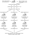
Figure 1
Figure 1
Schematic representation of the selection for B. burgdorferi proteins that mediate interactions with endothelial cells in vivo.
Figure 2
Figure 2
Alignment of P66 sequences selected in vivo with the B. burgdorferi strain B31 M1 structural gene sequence, amino acids 61-376. The two clones, F364 and K1176, were obtained from different starting library pools and from different tissues. The portion of P66 identified in an in vitro selection for β3 integrin binding is underlined, the peptide that inhibits B. burgdorferi attachment to β3 integrins is highlighted. Dashes indicate absence of amino acids corresponding to those in the strain B31 M1 protein.
Figure 3
Figure 3
The in vivo-selected portion of OspC. Panel A: the selected sequence aligned to two different sequences previously deposited in the database for B. burgdorferi strain N40 (Bunikis et al., 2004, Shih & Chao, 2002), and to the B31 sequence (http://www.tigr.org) (Fraser et al., 1997, Casjens et al., 2000). “fd” denotes the sequence common to our selected clones. Panel B: The regions selected in vivo are, in order on the polypeptide chain, loop 2 (green), beta sheet 2 (blue), loop 3 (green), alpha helix 2 (red), loop 4 (green), alpha helix 3 (red), loop 5 (green), and alpha helix 4 (red). The regions shaded gray (alpha helices 1 and 5, beta sheet 1, and loop 6) were not selected in vivo. For the sake of clarity, the two monomers have been separated and the side chains are not shown in the non-selected (gray) regions. The image was created using the structural data deposited by Eicken et al. (Eicken et al., 2001) in the Protein Data bank, Research Collaboratory for Structural Bioinformatics, Rutgers University (http://www.rcsb.org/pdb/Welcome.do), code 1G5Z, and customized for this figure using Swiss Pdb Viewer (http://www.expasy.ch/spdbv/mainpage.html).
Figure 3
Figure 3
The in vivo-selected portion of OspC. Panel A: the selected sequence aligned to two different sequences previously deposited in the database for B. burgdorferi strain N40 (Bunikis et al., 2004, Shih & Chao, 2002), and to the B31 sequence (http://www.tigr.org) (Fraser et al., 1997, Casjens et al., 2000). “fd” denotes the sequence common to our selected clones. Panel B: The regions selected in vivo are, in order on the polypeptide chain, loop 2 (green), beta sheet 2 (blue), loop 3 (green), alpha helix 2 (red), loop 4 (green), alpha helix 3 (red), loop 5 (green), and alpha helix 4 (red). The regions shaded gray (alpha helices 1 and 5, beta sheet 1, and loop 6) were not selected in vivo. For the sake of clarity, the two monomers have been separated and the side chains are not shown in the non-selected (gray) regions. The image was created using the structural data deposited by Eicken et al. (Eicken et al., 2001) in the Protein Data bank, Research Collaboratory for Structural Bioinformatics, Rutgers University (http://www.rcsb.org/pdb/Welcome.do), code 1G5Z, and customized for this figure using Swiss Pdb Viewer (http://www.expasy.ch/spdbv/mainpage.html).
Figure 4
Figure 4
Binding of selected phage clones to mammalian cells in culture. Individual phage clones containing the common selected B. burgdorferi sequences were added to confluent monolayers of cells, or to wells in which the cell culture medium, but no cells, had been plated. Phage binding was quantified by ELISA using an anti-M13 antibody; the inoculum was measured in separate wells in which phage were immediately fixed. Shown are the means + standard deviations of four replicates from a single representative experiment, after subtraction of binding to wells without cells. Each phage clone was tested for binding to at least 6 cell lines in at least four independent experiments.
Figure 5
Figure 5
Binding of ErpK and OspC to mammalian cell lines. The cells were allowed to grow to 90–100% confluence in 96 well plates. Control wells contained the cell culture medium but no cells. Recombinant MBP fusion proteins were added at the concentrations shown and incubated for 1.5 hours. Unbound proteins were removed by washing, then the wells were fixed. Binding of each MBP fusion and the control MBP alone was quantified by ELISA using a commercially available anti-MBP antiserum. Shown are the means ± standard deviations of 4 replicates from one of several (4–6) independent experiments for each panel. Data are shown for a cell line to which a corresponding phage clone binds efficiently.
Figure 6
Figure 6
Titers of individual phage clones in mouse tissues. Individual representative phage clones containing the common B. burgdorferi sequences selected in vivo were injected through the tail vein into mice and allowed to circulate. After perfusion of the mouse circulation through the heart, the tissues were harvested, weighed, homogenized, and diluted serially 10−1 to 10−8. Each dilution was used to infect E. coli, and resulting colonies were enumerated the following day. Shown are the log transducing units (TU)/gm tissue normalized to the input phage titers for at least three independent experiments for each clone. P66 bound significantly more efficiently than the vector control phage fdDog with one star denoting a p value < 0.05 and two stars indicating a p value < 0.01. Vls and ErpK show statistically significant differences compared to fdDog in the ear, with p values of 0.0176 and 0.0364, respectively. For Vls the p values for heart and tibiotarsus are 0.08, and for bladder 0.06. Note that the value plotted do not take into account 23 fold greater efficiency of fdDog over the library phage in infection of E. coli cells (unpublished observation).
Figure 7
Figure 7
Analysis of tissue-selected phage pools by quantitative PCR. Plasmid DNA (the replicative form of the phage genome) was purified from each tissue-selected pool, and genes of interest were amplified using real-time PCR. The means and standard deviations of 6–8 replicate experiments, each done in triplicate, are shown after conversion of Ct to copy number using standard curves consisting of phage genomic DNA. Although dbpA was not detected in the selected pools, it was easily detectable in the unselected library.

Similar articles

Cited by

References

    1. Akins DR, Porcella SF, Popova TG, Shevchenko D, Baker SI, Li M, Norgard MV, Radolf JD. Evidence for in vivo but not in vitro expression of a Borrelia burgdorferi outer surface protein F (OspF) homologue. Molecular microbiology. 1995;18:507–520. - PubMed
    1. Alitalo A, Meri T, Lankinen H, Seppala I, Lahdenne P, Hefty PS, Akins D, Meri S. Complement inhibitor factor H binding to Lyme disease spirochetes is mediated by inducible expression of multiple plasmid-encoded outer surface protein E paralogs. J Immunol. 2002;169:3847–3853. - PubMed
    1. Anguita J, Ramamoorthi N, Hovius JW, Das S, Thomas V, Persinski R, Conze D, Askenase PW, Rincon M, Kantor FS, Fikrig E. Salp15, an ixodes scapularis salivary protein, inhibits CD4+ T cell activation. Immunity. 2002;16:849–859. - PubMed
    1. Arap W, Kolonin MG, Trepel M, Lahdenranta J, Cardo-Vila M, Giordano RJ, Mintz PJ, Ardelt PU, Yao VJ, Vidal CI, Chen L, Flamm A, Valtanen H, Weavind LM, Hicks ME, Pollock RE, Botz GH, Bucana CD, Koivunen E, Cahill D, Troncoso P, Baggerly KA, Pentz RD, Do KA, Logothetis CJ, Pasqualini R. Steps toward mapping the human vasculature by phage display. Nat Med. 2002;8:121–127. - PubMed
    1. Arap W, Pasqualini R, Ruoslahti E. Cancer treatment by targeted drug delivery to tumor vasculature in a mouse model. Science. 1998;279:377–380. - PubMed

Publication types