. 2007 Sep 4:8:40.
doi: 10.1186/1471-2121-8-40.

3D cell nuclei segmentation based on gradient flow tracking

Affiliations

3D cell nuclei segmentation based on gradient flow tracking

Gang Li et al. BMC Cell Biol. .

Abstract

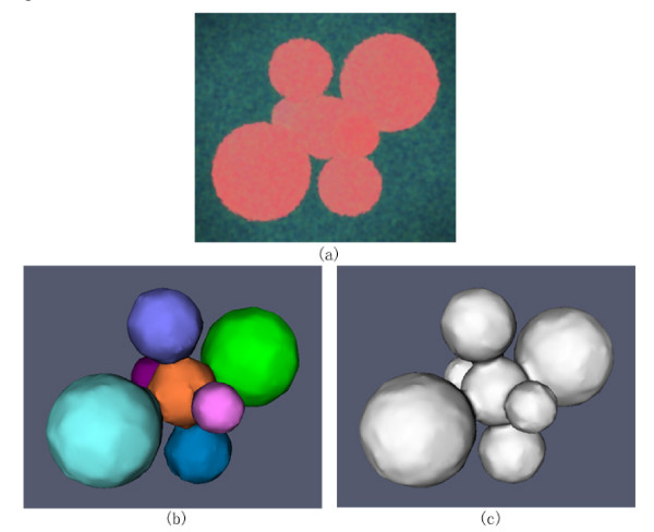
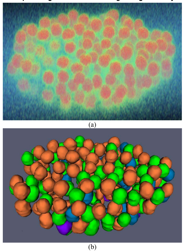
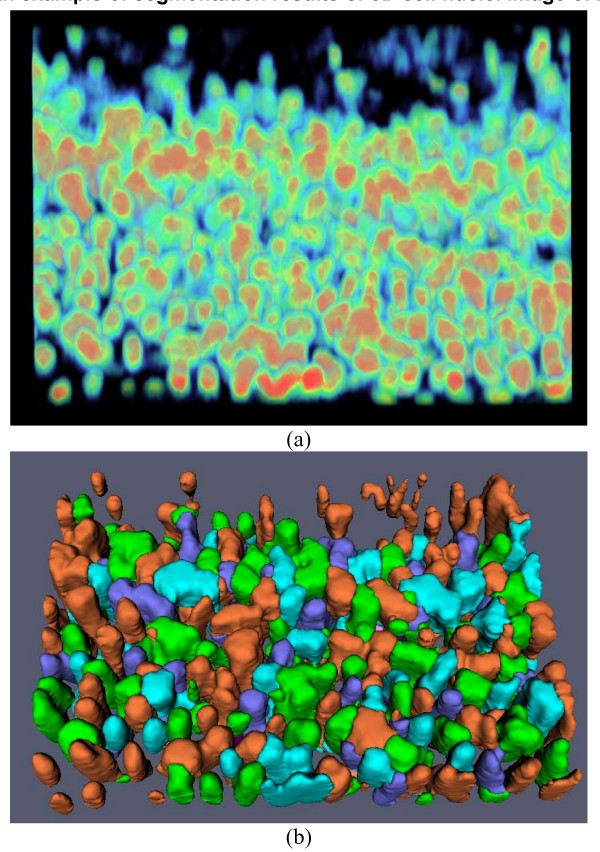
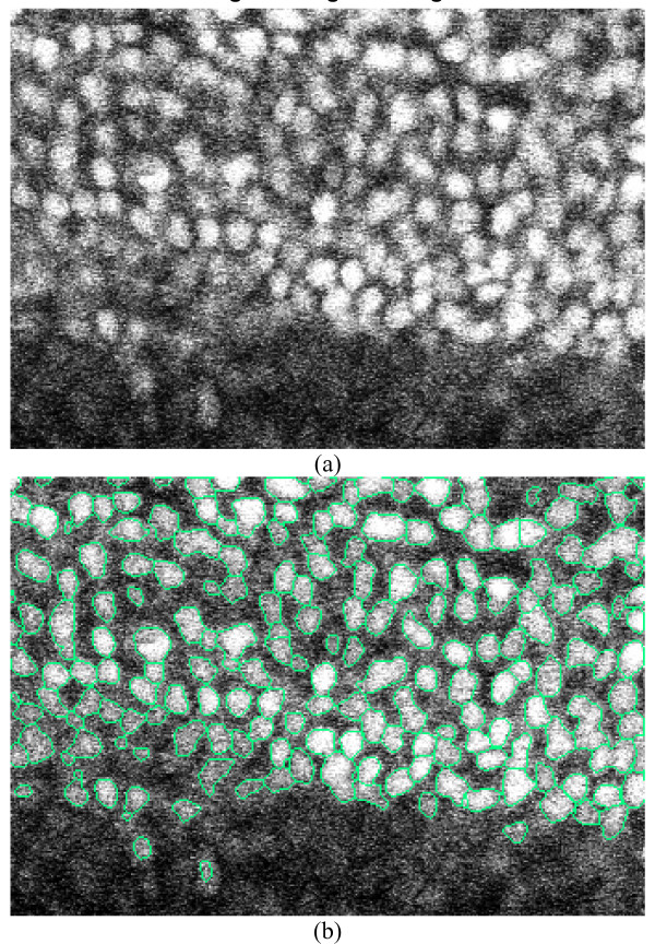
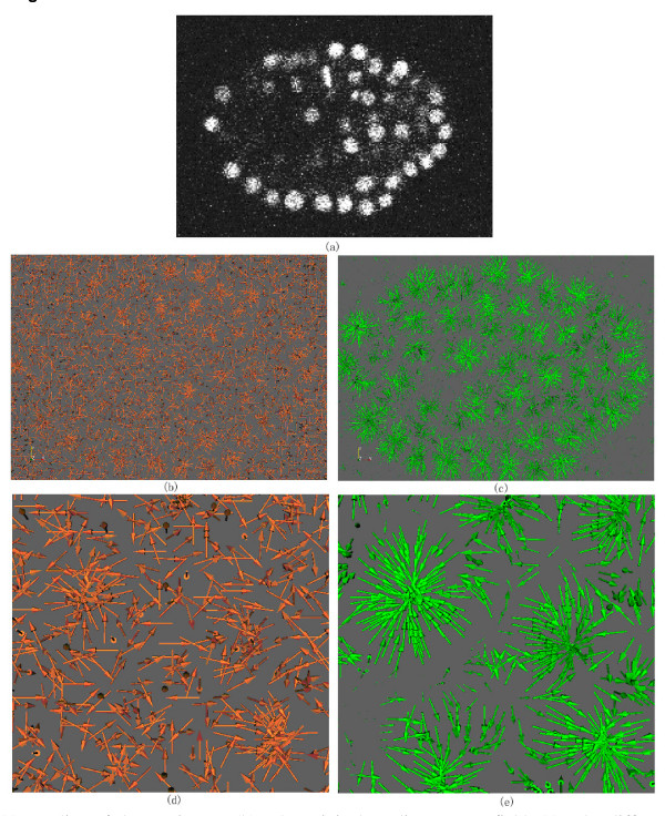
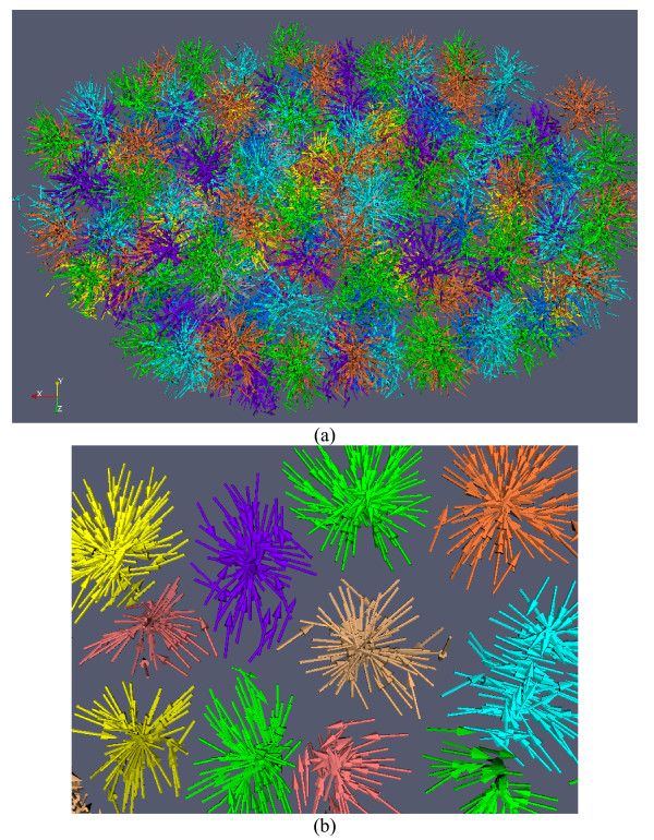
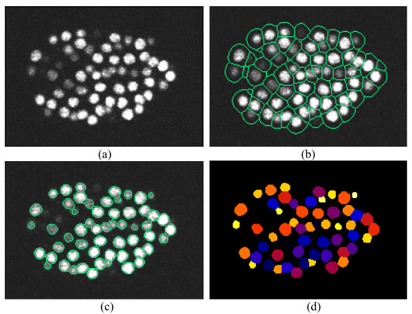
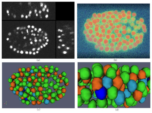
Background: Reliable segmentation of cell nuclei from three dimensional (3D) microscopic images is an important task in many biological studies. We present a novel, fully automated method for the segmentation of cell nuclei from 3D microscopic images. It was designed specifically to segment nuclei in images where the nuclei are closely juxtaposed or touching each other. The segmentation approach has three stages: 1) a gradient diffusion procedure, 2) gradient flow tracking and grouping, and 3) local adaptive thresholding.

Results: Both qualitative and quantitative results on synthesized and original 3D images are provided to demonstrate the performance and generality of the proposed method. Both the over-segmentation and under-segmentation percentages of the proposed method are around 5%. The volume overlap, compared to expert manual segmentation, is consistently over 90%.

Conclusion: The proposed algorithm is able to segment closely juxtaposed or touching cell nuclei obtained from 3D microscopy imaging with reasonable accuracy.


Figures

Figure 5
Figure 5
Result on synthesized 3D image segmentation. (a) The cross-sectional views of binary mask of the synthesized image. (b) The cross-sectional views of synthesized image with added noise. (c) The cross-sectional views of the overlaid boundaries of segmentation on synthesized noisy 3D image. (d) The cross-sectional views of randomly color-coded segmentation result.
Figure 6
Figure 6
Volume and surface rendering of synthesized 3D cell nuclei image and segmentation results. (a) Volume rendering of the synthesized noisy image. (b) Surface rendering of the segmentation results with the proposed method, in which each color indicates a segmented object. (c) Surface rendering of the segmentation results with the global Otsu thresholding, in which the touching objects are not divided correctly.
Figure 7
Figure 7
An example of segmentation result using C. elegans embryo image. (a) Volume rendering of original 3D image. (b) Surface rendering of segmentation results. For the convenience of inspection of touching cell nuclei, the results are randomly color-coded.
Figure 8
Figure 8
An example of segmentation results of 3D cell nuclei image of zebrafish. (a) Volume rendering of original 3D image. (b) Surface rendering of segmentation results. For the convenience of inspection of touching cell nuclei, the results are randomly color-coded.
Figure 9
Figure 9
2D slice view of the original image and segmentation result in Figure 8. (a). Original image. (b). The segmented image in which the green curves represent the boundaries of the cell nuclei.
Figure 1
Figure 1
The 3D view of gradient vector field and diffused gradient vector field with elastic deformation transformation of a slice cropped from a 3D cell nuclei image. (a). A slice of the 3D image. (b). The original gradient vector field. (c). The diffused gradient flow field with elastic deformation transformation. Obviously, the diffused vector field with elastic deformation transformation smoothly flows toward the central areas of cell nuclei. (d). Zoomed view of (b). (e). Zoomed view of (c).
Figure 2
Figure 2
Illustration of the gradient flow tracking based segmentation method. (a). Clustered gradient vectors in 3D space. (b). Zoomed view of selected clusters of gradient vectors. Each color represents a separated cell.
Figure 3
Figure 3
Illustration of 3D cell nuclei segmentation on a 2D slice. (a) A slice from 3D cell nuclei image. (b)Boundaries of small regions overlaid on the slice. The boundaries are obtained by the method in Section 2.2. (c) Edges of cell nuclei overlaid on the slice after the step of adaptive thresholding in Section 2.3. (d) Randomly color-coded extracted cells.
Figure 4
Figure 4
Illustration of cell nuclei segmentation in 3D space. (a). Cross-sectional views of a 3D cell nuclei image. (b) Volume rendering of 3D cell nuclei image. (c) Surface rendering of extracted 3D cell nuclei. For the convenience of inspection of touching cell nuclei, the results are randomly color-coded. (d) Zooming view of the results.

References

    1. Holley SA, Geisler R, Nüsslein-Volhard C. Control of her1 expression during zebrafish somitogenesis by a Delta-dependent oscillator and an independent wavefront activity. Genes & Dev. 2000;14:1678–1690. - PMC - PubMed
    1. Jülich D, Hwee LC, Round J, Nicoliaje C, Scroeder J, Davies A, Geisler R, Lewis J, Jiang YJ, Holley SA. beamter/deltaC and the role of Notch ligands in the zebrafish somite segmentation, hindbrain neurogenesis and hypochord differentiation. Dev Biol. 2005;286:391–404. doi: 10.1016/j.ydbio.2005.06.040. - DOI - PubMed
    1. Lin G, Adiga U, Olson K, Guzowski J, Barnes C, Roysam B. A hybrid 3-D watershed algorithm incorporating gradient cues and object models for automatic segmentation of nuclei in confocal image stacks. Cytometry. 2003;56A:23–36. doi: 10.1002/cyto.a.10079. - DOI - PubMed
    1. Lin G, Chawla MK, Olson K, Guzowski JF, Barnes CA, Roysam B. Hierarchical, model-based merging of multiple fragments for improvoed three-dimensional segmentation of nuclei. Cytometry. 2005;63A:20–33. doi: 10.1002/cyto.a.20099. - DOI - PubMed
    1. Umesh Adiga PS, Chaudhuri BB. An efficient method based on watershed and rule-based merging for segmentation of 3-D histo-pathological images. Pattern Recognition. 2001;34:1449–1458. doi: 10.1016/S0031-3203(00)00076-5. - DOI

Publication types

MeSH terms