. 2007 Nov;27(21):7381-93.
doi: 10.1128/MCB.00440-07. Epub 2007 Sep 4.

Hypoxia-inducible factor 1 and dysregulated c-Myc cooperatively induce vascular endothelial growth factor and metabolic switches hexokinase 2 and pyruvate dehydrogenase kinase 1

Affiliations

Hypoxia-inducible factor 1 and dysregulated c-Myc cooperatively induce vascular endothelial growth factor and metabolic switches hexokinase 2 and pyruvate dehydrogenase kinase 1

Jung-whan Kim et al. Mol Cell Biol. 2007 Nov.

Abstract

Hypoxia is a pervasive microenvironmental factor that affects normal development as well as tumor progression. In most normal cells, hypoxia stabilizes hypoxia-inducible transcription factors (HIFs), particularly HIF-1, which activates genes involved in anaerobic metabolism and angiogenesis. As hypoxia signals a cellular deprivation state, HIF-1 has also been reported to counter the activity of MYC, which encodes a transcription factor that drives cell growth and proliferation. Since many human cancers express dysregulated MYC, we sought to determine whether HIF-1 would in fact collaborate with dysregulated MYC rather countering its function. Here, using the P493-6 Burkitt's lymphoma model with an inducible MYC, we demonstrate that HIF-1 cooperates with dysregulated c-Myc to promote glycolysis by induction of hexokinase 2, which catalyzes the first step of glycolysis, and pyruvate dehydrogenase kinase 1, which inactivates pyruvate dehydrogenase and diminishes mitochondrial respiration. We also found the collaborative induction of vascular endothelial growth factor (VEGF) by HIF-1 and dysregulated c-Myc. This study reports the previously unsuspected collaboration between HIF-1 and dysregulated MYC and thereby provides additional insights into the regulation of VEGF and the Warburg effect, which describes the propensity for cancer cells to convert glucose to lactate.


Figures

FIG. 1.
FIG. 1.
Effects of constitutively stable HIF-1α (CA5-HIF-1α) on tumorigenesis and expression of HK2 and PDK1. (A) Tumor growth of P493-6 cells expressing CA5-HIF-1α or control EV with or without the tetracycline analog doxycycline (0.01%, wt/vol) treatment through drinking water. Tumor volumes were determined by caliper measurements. Error bars represent standard errors of the means (n = 20 for non-tetracycline-treated tumors, n = 10 for tetracycline-treated tumors). (B) Immunoblot assay (top) and its quantification (bottom) of lysates from xenografts of P493-6 cells expressing CA5-HIF-1α or control EV. The average optical density from two bands for each CA5-HIF-1α and EV control P493-6 xenograft is represented. Error bars represent ranges of two bands. (C) Immunoblot assay (top) and its quantification of induction (n-fold) at 48 h (bottom) of HK2, PDK1, LDHA, HK1, CA5-HIF-1α, and c-Myc in CA5-HIF-1α or control EV P493-6 cells after withdrawal of tetracycline under nonhypoxic conditions. Actin is shown as a loading control. *, P < 0.02 by Student's t test.
FIG. 2.
FIG. 2.
Enhanced glycolysis by c-Myc and HIF-1 in P493-6 cells. (A) Immunoblot assays of c-Myc and HIF-1α expression in P493-6 cells. P493-6 cells were pretreated with tetracycline (0.1 μg/ml) for 48 h. After washing, cells were cultured with or without tetracycline in hypoxia (0.1% O2) or normoxia as indicated in the bottom. To induce HIF-1α in nonhypoxic conditions, P493-6 cells were incubated in medium containing 100 μM CoCl2. Equal amounts of nuclear protein were subjected to immunoblot assay to determine the expressions of c-Myc and HIF-1α. Topoisomerase 1 is shown as a loading control for nuclear extracts. (B) Cellular glucose uptake in indicated conditions at 48 h was determined by measuring intracellular 2-[3H]deoxyglucose. Error bars represent standard errors of the means from two independent experiments. (C) Lactate accumulation in the medium was measured using a lactate assay kit. Lactate concentrations were normalized to viable cell number. Error bars represent standard errors of the means from two independent experiments.
FIG. 3.
FIG. 3.
Enhanced HK2 expression by c-Myc and HIF-1. (A) Northern blot analysis of HK2 expression in P493-6 cells. Pretreatment with tetracycline for 48 h to suppress MYC expression is followed by incubating cells in different conditions as illustrated. Ethidium bromide staining of 18S is shown as a loading control. (B) Immunoblot assays of HK2 expression in P493-6 cells. Actin is shown as a loading control. (C) HK enzymatic activities. Total cell lysates from P493-6 cells cultured with or without tetracycline in hypoxia (left) or CoCl2 treatment (right) for 29 h were subjected to HK activity assay. HK activity is determined by NADH production at 340 nm and normalized by total protein concentration. Error bars represent standard deviations from three independent experiments.
FIG. 4.
FIG. 4.
Effect of decreased HIF-1α expression on induction of HK2 and PDK1. (A) Immunoblot of HIF-1α and c-Myc expression in P493-6 cells stably expressing shRNA targeting HIF-1α or control vector. HIF-1α shRNA or control P493-6 cells were pretreated with tetracycline (0.1 μg/ml) for 48 h. After washing, cells were cultured in hypoxia (0.1% O2). Actin is shown as a loading control. (B) Immunoblot of HK2, PDK1, and p21 expression. HIF-1α shRNA or control P493-6 cells were pretreated with tetracycline (0.1 μg/ml) for 48 h. After washing, cells were cultured with or without tetracycline in hypoxia (0.1% O2) or normoxia as indicated in the bottom. Actin is shown as a loading control.
FIG. 5.
FIG. 5.
Effect of decreased HK2 expression on glycolysis in P493-6 cells. (A) Immunoblot assays of HK2 expression in P493-6 cells electroporated with pools of four different siRNAs targeting HK2 (siGENOME SMART POOL) or control scrambled siRNA in hypoxia (0.1% O2). HK1 is shown as a loading control. (B and C) Glucose uptake (B) and lactate accumulation (C) of P493-6 cells. Cells in indicated conditions at 48 h were subjected to measurement of intracellular 2-[3H]deoxyglucose or lactate accumulation in the culture medium. Protein concentration from viable cells or total number of viable cells is used for normalization of glucose uptake or lactate accumulation, respectively. Error bars represent standard errors of the means from two or three independent experiments.
FIG. 6.
FIG. 6.
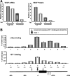
Scanning ChIP assay of the HK2 gene. The human HK2 gene was scanned by ChIP to determine the c-Myc (top panel) or HIF-1 (middle panel) DNA binding regions. Labeled regions (amplicons 1 to 22) of the HK2 gene were quantitatively measured by real-time PCR. PCR was performed on the fragmented chromatin precipitated from non-tetracycline-treated hypoxic (72-h) P493-6 cells with anti-c-Myc or anti-HIF-1α, without antibody, or control IgG antibody (see Fig. S2 in the supplemental material for the background signals of no-antibody and IgG antibody controls). Binding activity is represented as a percentage of total input of the chromatin DNA. The bottom panel indicates the locations of conserved canonical E boxes (large arrows), conserved HIF-1 binding sites (small arrows), and exon 1 in the human HK2 genomic sequence. The amplicons for scanning ChIP are indicated by the lines above the HK2 gene and labeled. Error bars represent standard deviations from two or three independent duplicate reactions.
FIG. 7.
FIG. 7.
Enhanced PDK1 expression by c-Myc and HIF-1 in P493-6 cells. (A) mRNA levels of PDK1 are determined by real-time RT-PCR. Error bars represent standard deviations from two independent duplicate reactions. (B) Immunoblot assays of PDK1 expression in hypoxic (or CoCl2 treated) or nonhypoxic P493-6 cells that were treated with or without tetracycline. Actin is shown as a loading control. (C) Immunoblot assay of decreased PDK1 expression by PDK1 siRNA or control siRNA in P493-6 cells. CoCl2 treatment was used to induce HIF-1-dependent PDK1 expression. Actin is shown as a loading control. (D and E) Lactate accumulation in the culture medium of P493-6 cells with or without CoCl2 treatment to induce PDK1 expression. PDK1 is inhibited by treatment with either siRNA (D) or a PDK inhibitor, dichloroacetate (DCA) (E). Lactate concentrations are normalized to total viable cells. Error bars represent standard errors of the means from two to three independent experiments.
FIG. 8.
FIG. 8.
Scanning ChIP assay of the PDK1 gene. Labeled regions (amplicons 1 to 9) of the PDK1 gene were quantitatively amplified by real-time PCR. Error bars represent standard deviations from two or three independent duplicate reactions. Symbols are identical to those in Fig. 6.
FIG. 9.
FIG. 9.
Enhanced VEGF expression by c-Myc and HIF-1. (A) mRNA and protein levels of VEGF were determined by real-time RT-PCR and ELISA, respectively. Error bars represent standard deviations from two independent duplicate reactions. (B) DNA binding of c-Myc and HIF-1 to the VEGF gene was determined by scanning ChIP assay as described for Fig. 6.

Similar articles

Cited by

References

    1. Adhikary, S., and M. Eilers. 2005. Transcriptional regulation and transformation by Myc proteins. Nat. Rev. Mol. Cell Biol. 6: 635-645. - PubMed
    1. Ashrafian, H. 2006. Cancer's sweet tooth: the Janus effect of glucose metabolism in tumorigenesis. Lancet 367: 618-621. - PubMed
    1. Baudino, T. A., C. McKay, H. Pendeville-Samain, J. A. Nilsson, K. H. Maclean, E. L. White, A. C. Davis, J. N. Ihle, and J. L. Cleveland. 2002. c-Myc is essential for vasculogenesis and angiogenesis during development and tumor progression. Genes Dev. 16: 2530-2543. - PMC - PubMed
    1. Berra, E., E. Benizri, A. Ginouves, V. Volmat, D. Roux, and J. Pouyssegur. 2003. HIF prolyl-hydroxylase 2 is the key oxygen sensor setting low steady-state levels of HIF-1α in normoxia. EMBO J. 22: 4082-4090. - PMC - PubMed
    1. Bonnet, S., S. L. Archer, J. Allalunis-Turner, A. Haromy, C. Beaulieu, R. Thompson, C. T. Lee, G. D. Lopaschuk, L. Puttagunta, G. Harry, K. Hashimoto, C. J. Porter, M. A. Andrade, B. Thebaud, and E. D. Michelakis. 2007. A mitochondria-k(+) channel axis is suppressed in cancer and its normalization promotes apoptosis and inhibits cancer growth. Cancer Cell 11: 37-51. - PubMed

Publication types

MeSH terms